Skip to main content
Search
Search Toggle Button
Select your language
United States
América Latina
Australia
Brasil
Canada
Canada (Français)
Deutschland
España
France
India
Italia
United Kingdom
Россия
简体中文
Menu
Main menu
Products
Ultrasound systems
Sonosite LX
Unmatched frequency range
Sonosite ST
Procedural Partner
Image
Sonosite MT
New
Built to Move
Sonosite PX
Flexible hybrid
Discover Products
Sonosite PIV Assist
New
Sonosite UHF 46-20
New
Transducers
Accessories
Sonosite Voice Assist
Sonosite Synchronicity
Education
Learn
Clinical Images & Videos
Conferences & Society Meetings
Sonosite Institute
Webinars
Educational Resources
Support
Biomed Service Training
Cleaners and Disinfectants
Connectivity
Contact Support
Dealers & Support Staff
FAQ
Product Retirement
Security
Symbols Glossary
User Documents
Warranty and Service
About
Why Sonosite
Global Health Programme
Leadership Team
Press Releases
What is POCUS?
Careers
Blog
Contact Us
POCUS 101
Learn Ultrasound Basics
Search
Clinical Specialties
- Any -
Admin
Anaesthesiology
BioMed
Cardiology
Cath Lab
Chiropractic
Clinical Educator
Dermatology
EMED
EMS/Air Med
Endocrinology
FP/GP
Gastroenterology
Global Health
Government
Home Health
ICU/CCU
Informatics
Internal Medicine
Interv Rad
Medical Education
MFM/Perinatology
Nephrology
Neurology
Nursing
OB/GYN
Oncology
Opthamology
OR (Shared System)
Orthopedics
Osteopathic Medicine
Pain Mgmt
Paediatrics
Physical Med & Rehab
Physical Therapy
Podiatry
Pulmonary
Purchasing
Radiology
Respiratory
Rheumatology
SIM Center
Sports Medicine
Sports Team
Surgery
Surgery (Breast)
Urgent Care
Urology
Vascular
Vascular Lab
Vascular Surgery
Vein Clinic
Type
- Any -
Case Study Videos
How To Videos
Other Videos & Images
Image
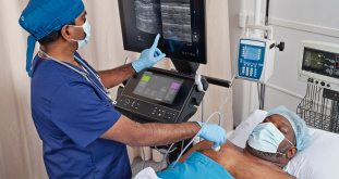
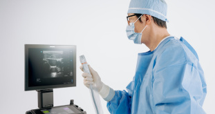
Sonosite Phased Array Ultrasound Transducers: Clinical Applications and Key Advantages
16132
Image
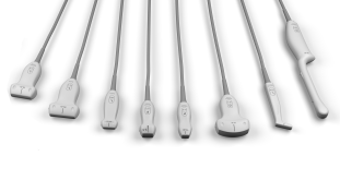
Understanding the Basics of Ultrasound Probe Types
16064
Image
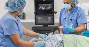
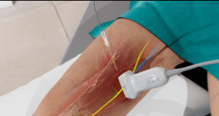
How to Perform an Ultrasound-Guided Supraclavicular Brachial Plexus Block
15956
Image
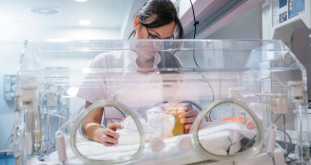
5 Steps to Setting Up a Point-of-Care Ultrasound Programme in the Neonatal Intensive Care Unit
15928
Image
What is Ultra High Frequency Ultrasound?
15926
Image
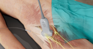
How to perform a Lateral Femoral Cutaneous Nerve Block
15868
Image
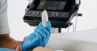
How to Choose the Right Portable Ultrasound Transducer
15858
Image
How to Perform an Ultrasound-Guided PENG Block
15838
Image
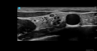
Metacarpal Head Cartilage Damage in Rheumatoid Arthritis
15836
Image
The Use of Ultra-High Frequency Ultrasound for Vascular Access in Paediatric Patients
15830
Image
How to Choose the Best Portable Ultrasound Machine for Your Practise
15640
Image
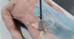
How to Perform a Rectus Sheath Block
15638
Image
How to Perform an IPACK Block with Ultrasound
15636
Image
How to Perform a Popliteal Sciatic Nerve Block
15614
Image
How to Perform an Adductor Canal Block
15610
Image
An Introduction to VExUS - Venous Excess Ultrasound Exam
15596
Current page
1
Page
2
Page
3
Page
4
Page
5
Page
6
Page
7
Page
8
Page
9
…
Next page
››
Last page
Last »
Products
Discover Products
Sonosite MT
Sonosite LX
Sonosite PX
Sonosite ST
Sonosite PIV Assist
Sonosite UHF 46-20
Transducers
Accessories
Sonosite Voice Assist
Sonosite Synchronicity
Sonosite LX
Education
Learn
Clinical Images & Videos
Conferences & Society Meetings
Sonosite Institute
Webinars
Educational Resources
Support
Biomed Service Training
Cleaners and Disinfectants
Connectivity
Contact Support
Dealers & Support Staff
FAQ
Product Retirement
Security
Symbols Glossary
User Documents
Warranty and Service
About
Why Sonosite
Global Health Programme
Leadership Team
Press Releases
What is POCUS?
Careers
Blog
Contact Us
Select your language
United States
América Latina
Australia
Brasil
Canada
Canada (Français)
Deutschland
España
France
India
Italia
United Kingdom
Россия
简体中文