

Image
Objective:
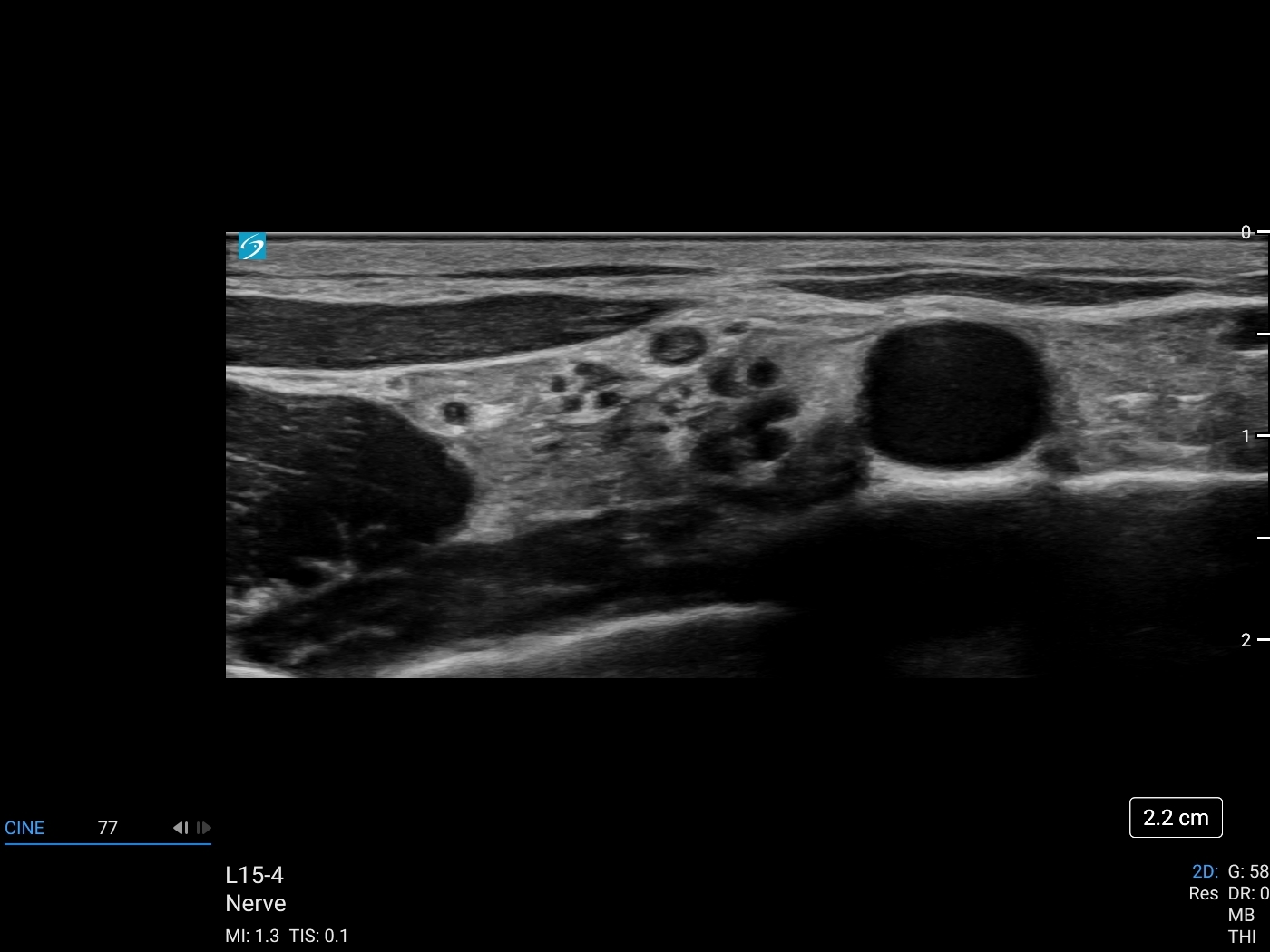
Spread of local anaesthetic surrounding the brachial plexus lateral to the subclavian artery and superior to the 1st rib.
Technique:
- The ultrasound transducer is placed transversely, mid-clavicle within the supraclavicular fossa.
- The brachial plexus is located lateral to the subclavian artery and superior to the first rib.
- Ideal ultrasound image of the supraclavicular brachial plexus should include: First rib, subclavian artery, and pleura. A clear delineation between the artery and brachial plexus should be visualised.
- In-plane needle insertion with a lateral to medial approach.
- Colour Doppler can be utilised to confirm subclavian artery position along with identification of any vessel anatomical variations.
Patient Positioning:
Supine
Transducers:
Click to download the guide today.