

Point-of-care ultrasound (POCUS) relies on more than just the ultrasound system itself. The probe you choose plays a critical role in image quality, diagnostic accuracy, and clinical confidence. With different ultrasound probe types designed for specific depths, frequencies, and clinical applications, understanding how each transducer works is foundational to using ultrasound effectively at the bedside. This article breaks down the basics of POCUS probe types, explaining how they differ, when to use them, and why selecting the right probe matters across a wide range of clinical scenarios.
Sonosite's Ultrasound Probe Types and Applications:
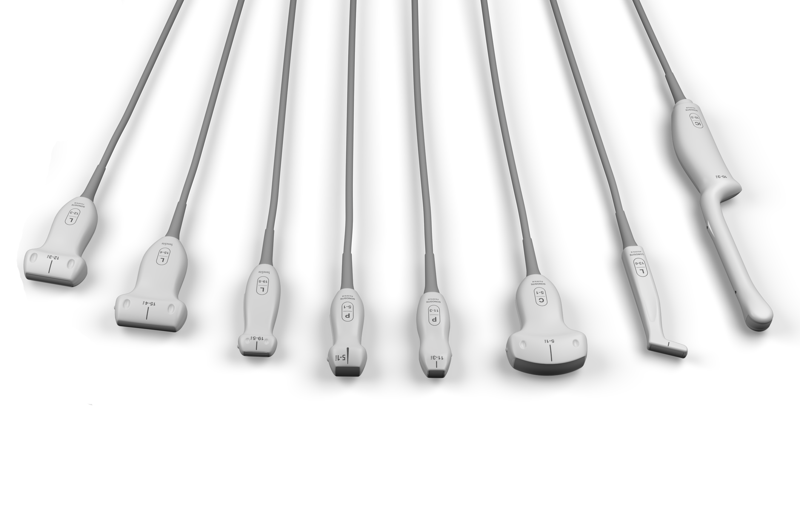
FUJIFILM Sonosite ultrasound probes vary in design to meet specific clinical needs, each excelling in particular areas of patient care:
Linear Probes:
- Features: A large range of frequencies (up to 46MHz) for superb resolution of shallow structures.
- Applications: Vascular access, soft tissue evaluations, musculoskeletal imaging, central line placements, and superficial nerve localization.
Curvilinear (Convex) Probes:
- Features: Low to mid-frequency (1–10 MHz) providing deeper penetration.
- Applications: Abdominal, pelvic, obstetric, liver, and bowel imaging, including trauma and gallbladder exams.
Phased Array Probes:
- Features: Small footprint, low-frequency (1–5 MHz) designed for deep targets.
- Applications: Cardiac, lung, and transcranial imaging, especially useful in challenging patient scenarios.
Endocavitary Probes:
- Features: Mid-frequency (5–8 MHz) curved probes designed for internal imaging.
- Applications: Transvaginal and transrectal scans for gynaecologic and urologic assessments; require high-level disinfection.
Key considerations for Choosing a POCUS Probe
Clinical Settings
Your choice of probes should align with your patient population and clinical focus:
- Emergency Medicine: Combination of phased array and curvilinear probes for trauma and cardiac, with linear probes for vascular and lung assessments.
- Paediatrics: High-frequency linear probes provide excellent resolution for small anatomy and vascular access.
- Critical Care: Mix of linear, curvilinear, and phased array probes accommodate vascular access, thoracentesis, echocardiography, and pleural exams.
- Musculoskeletal: High-frequency linear probes for joint injections and aspirations; curvilinear probes for deeper joints like hips.
Durability Matters
In high-turnover environments, probe durability is crucial. FUJIFILM Sonosite probes offer rugged, drop-tested designs with ergonomic features tailored for easy use and long-lasting performance, ideal for emergency rooms and intensive care units.
Cost vs. Long-Term Value
Investing in durable probes like those from FUJIFILM Sonosite pays off over time. Designed to withstand intensive use and repeated disinfection, these probes maintain performance and reliability under demanding clinical conditions. Their resistance to wear lowers replacement frequency and repair costs, ensuring efficient, cost-effective operations.
Conclusion: Choose the Right Probe for Your Practise
FUJIFILM Sonosite probes combine reliability, durability, and ergonomic design to optimise POCUS workflows across specialties. Selecting the ideal probe depends on your clinical focus, patient needs, and imaging demands. From emergency care to paediatrics and obstetrics, Sonosite offers a versatile, high-quality transducer portfolio designed to support clinicians in the delivery of patient-focused care.
Frequently Asked Questions About Ultrasound Probe Types
What are the main types of ultrasound probes?
The main types of ultrasound probes include linear, curvilinear (convex), phased array, and endocavitary probes. Each probe type is designed for specific imaging depths and clinical applications.
How does ultrasound probe frequency affect imaging?
Ultrasound probe frequency determines image resolution and depth of penetration. Higher-frequency probes provide better resolution for superficial structures, while lower-frequency probes allow imaging of deeper anatomy.
What is a linear ultrasound probe used for?
A linear ultrasound probe is used for imaging superficial structures such as blood vessels, nerves, soft tissue, and musculoskeletal anatomy. It typically operates at higher frequencies and produces high-resolution images.
What is a curvilinear ultrasound probe used for?
A curvilinear ultrasound probe is commonly used for abdominal, pelvic, and obstetric imaging. Its lower frequency allows for deeper penetration and a wider field of view compared to linear probes.
What is a phased array ultrasound probe?
A phased array ultrasound probe is a low-frequency probe with a small footprint, commonly used for cardiac and thoracic imaging. Its design allows imaging between ribs and visualisation of deep structures.
What are endocavitary ultrasound probes used for?
Endocavitary ultrasound probes are used for internal examinations such as transvaginal or transrectal imaging. Their design allows close proximity to internal organs for improved image quality.
Can one ultrasound probe be used for all exams?
No, a single ultrasound probe cannot be used for all exams. Different probe types are required based on imaging depth, target anatomy, and clinical application.
Why is ultrasound probe footprint important?
Ultrasound probe footprint affects probe placement and field of view. Smaller footprints are useful in tight anatomical spaces, while larger footprints provide broader anatomical visualisation.
How do clinicians choose the right ultrasound probe?
Clinicians choose an ultrasound probe based on the target anatomy, required imaging depth, and clinical use. Factors such as frequency, footprint, and field of view guide probe selection.
Are ultrasound probes specific to certain medical specialties?
Some ultrasound probes are optimised for specific medical specialties, such as cardiology or obstetrics, but many probes are versatile and used across multiple clinical settings.
Why is probe selection important in point-of-care ultrasound (POCUS)?
Probe selection is important in point-of-care ultrasound because it directly affects image quality, exam efficiency, and the clinician’s ability to accurately assess anatomy at the bedside.
*Please refer to product indications for use located in FUJIFILM Sonosite ultrasound system user guides.*