

Ultra-high frequency ultrasound represents a significant advancement in point-of-care imaging, enabling clinicians to visualise extremely small and superficial anatomical structures with remarkable clarity. Operating at frequencies far higher than conventional ultrasound, UHF ultrasound can produce ultra-detailed images of nerves, vessels, and soft tissues just beneath the skin, making it particularly valuable for applications such as vascular access, dermatology, musculoskeletal imaging, and paediatric care. As ultrasound technology continues to evolve, ultra-high frequency systems are expanding what clinicians can see and diagnose at the bedside, helping support more precise procedures and improved patient outcomes. This article presents frequently asked questions and common applications.
What is frequency?
In ultrasound, frequency refers to the number of sound wave cycles that occur in one second. It is measured in hertz (Hz). Ultrasound frequencies used in medical imaging typically range from 1 megahertz (MHz) to over 30 MHz, with higher frequencies providing better resolution but lower penetration depth, and lower frequencies allowing deeper penetration but with lower resolution. Ultrasound technology exploits this frequency range to provide detailed imaging of the body's internal structures, aiding clinicians in diagnostic and therapeutic procedures.
What are considered “high” or “ultra-high” frequencies?
Different ultrasound frequency ranges can meet different imaging requirements, balancing the need for resolution versus penetration depth. These frequency ranges help clinicians and technicians select the most appropriate ultrasound settings for various diagnostic and therapeutic needs, ensuring optimal image quality and diagnostic accuracy.
- High-Frequency Ultrasound (10-30 MHz): Used for imaging more superficial structures such as tendons, muscles, and organs close to the skin. These frequencies provide higher resolution images but do not penetrate as deeply.
- Ultra-High Frequency Ultrasound (30 MHz +): Useful for very high-resolution imaging of superficial structures, like small nerves, tendons, vessels, skin layers, and small joints, particularly in the first centimetre of the imaging field.
Why is ultra-high frequency special?
While ultrasound imaging technology has existed for use in diagnostic medicine since the 1950’s, there were technical barriers to accessing the upper range of frequencies. Dr. Stuart Foster, a researcher at Sunnybrook Research Instutute in Toronto, Canada through his research overcame these barriers and in 1999 founded what is now FUJIFILM VisualSonics, Inc which specialises in ultra-high frequency ultrasound equipment for preclinical research in the fields of cardiology, oncology, neurobiology, developmental biology and other fields. This led to the world’s first clinically available UHF ultrasound system, the Vevo MD, which enabled clinical researchers to explore the possibilities of these new frequency ranges for applications in surgical planning, dermatology, vascular biology and other application areas.
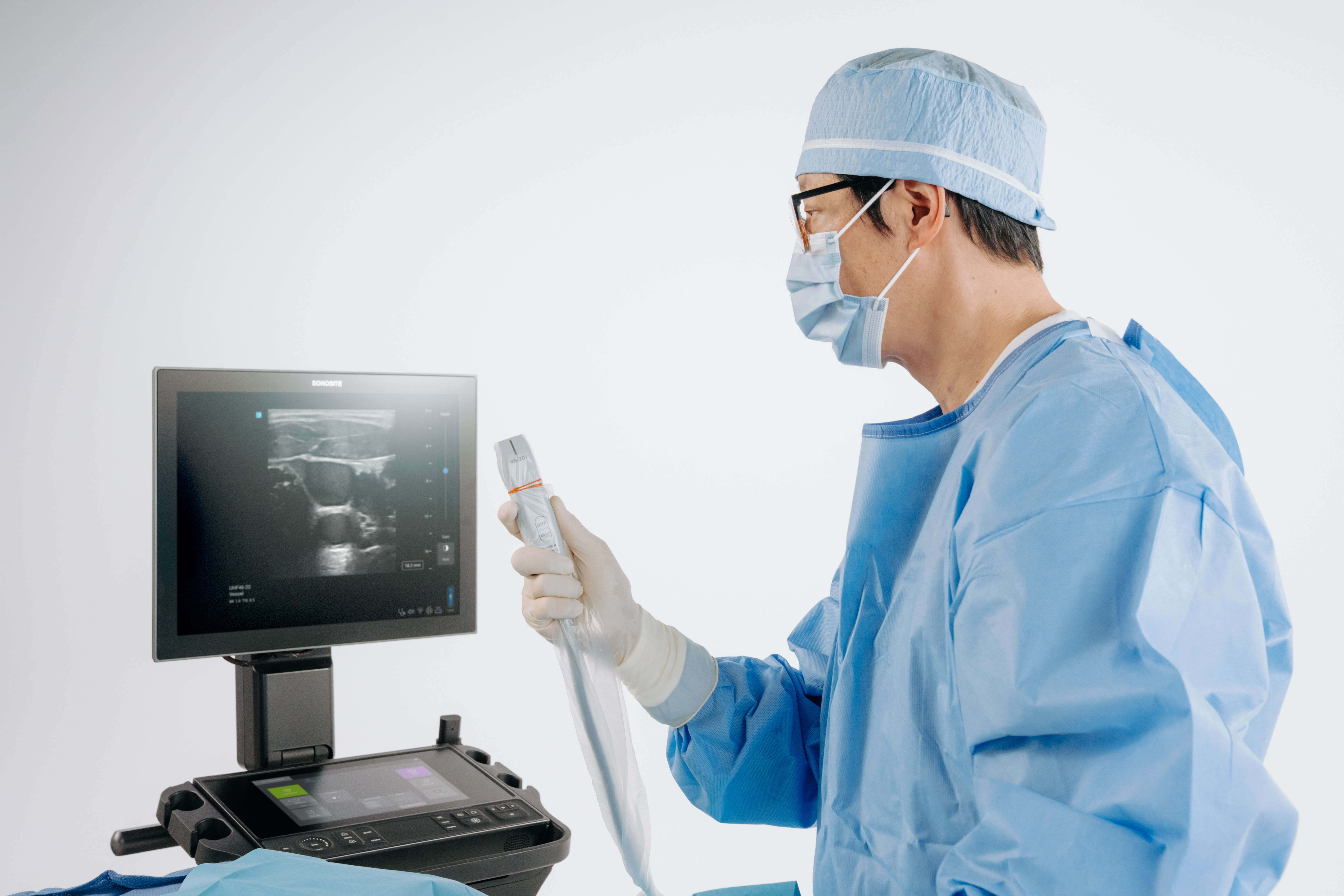
What does Sonosite offer?
Fujifilm Sonosite’s UHF Transducer is the first and only 46 MHz ultra-high frequency transducer available at the point of care.1
By partnering with Fujifilm VisualSonics, the industry leader in ultra-high frequency ultrasound imaging, Sonosite has now created the first point of care ultrasound system that can image both low and ultra-high frequencies. With UHF46-20, Sonosite LX now has the best superficial imaging, and the largest frequency range available on any POCUS system.1
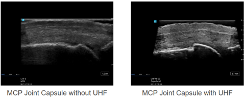
See detailed micro anatomy
Exceptional Microanatomy Visualisation: See superficial sub-millimetre vessels, nerves, and neonatal anatomy with unmatched clarity in point of care ultrasound, supporting more informed clinical decisions.2,3
Vascular access
Ultra-high frequency ultrasound may help clinicians improve procedural quality by allowing for improved visualisation of submillimetre superficial vessels2,4
Aid in Diagnosis and Intervention:
UHF may help you identify subclinical synovitis, erosions, crystal deposits, and inflammation, which may aid you in early diagnosis and intervention in chronic conditions.5,6,7
Conclusion
Advancements in ultrasound technology, particularly in ultra-high-frequency imaging, are expanding the capabilities of medical imaging and research. With tools like the Sonosite LX and the UHF46-20 transducer, clinicians and researchers have access to a broader range of frequencies, aiding in the visualisation of superficial structures with exceptional detail. These innovations support a variety of diagnostic and procedural applications, making ultrasound a versatile tool for both clinical and preclinical settings.
1. Internal Fujifilm research as of April 2025.
2. Latham, G. J., Veneracion, M. L., Joffe, D. C., Bosenberg, A. T., Flack, S. H., & Low, D. K. (2013). High-frequency micro-ultrasound for vascular access in young children--a feasibility study by the High-frequency UltraSound in Kids studY (HUSKY) group. Paediatric anaesthesia, 23(6), 529–535. https://doi.org/10.1111/pan.12131
3. Salvia, G.; Zerbinati, N.; Manzo Margiotta, F.; Michelucci, A.; Granieri, G.; Fidanzi, C.; Morganti, R.; Romanelli, M.; Dini, V. Ultra-High-Frequency Ultrasound as an Innovative Imaging Evaluation of Hyaluronic Acid Filler in Nasolabial Folds. Diagnostics 2023, 13, 2761. https://doi.org/10.3390/ diagnostics13172761
4. Brusciano, V., & Lecce, M. (2024). Advantages of the use of ultrasound in newborn vascular access: a systematic review. Journal of ultrasound, 27(2), 203–207. https://doi.org/10.1007/s40477-023-00832-1
5. Albano, D., Aringhieri, G., Messina, C., De Flaviis, L., & Sconfienza, L. M. (2020). High-Frequency and Ultra-High Frequency Ultrasound: Musculoskeletal Imaging up to 70 MHz. Seminars in musculoskeletal radiology, 24(2), 125–134. https://doi.org/10.1055/s-0039-3401042
6. Russo, A.; Reginelli, A.; Lacasella, G.V.; Grassi, E.; Karaboue, M.A.A.; Quarto, T.; Busetto, G.M.; Aliprandi, A.; Grassi, R.; Berritto, D. Clinical Application of Ultra-HighFrequency Ultrasound. J. Pers. Med. 2022, 12, 1733. https://doi.org/10.3390/jpm12101733
7. Ait Ichou, J., Gauvin, S., & Faingold, R. (2021). Ultra-high-frequency ultrasound of superficial and musculoskeletal structures in the paediatric population. Paediatric radiology, 51(9), 1748–1757. https://doi.org/10.1007/s00247-021-04978-0