

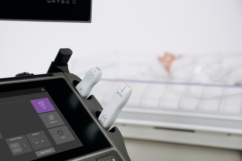
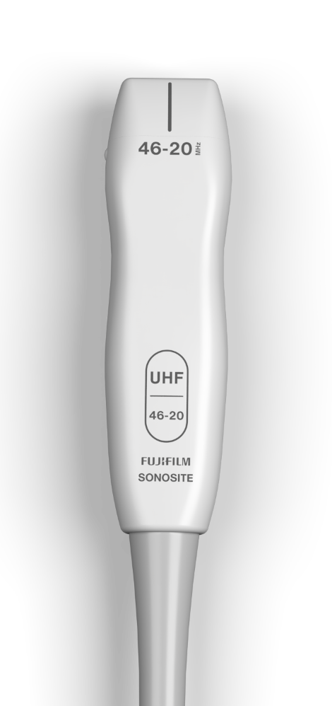
UHF 46-20 Ultrasound Transducer
How UHF46-20 transforms patient care
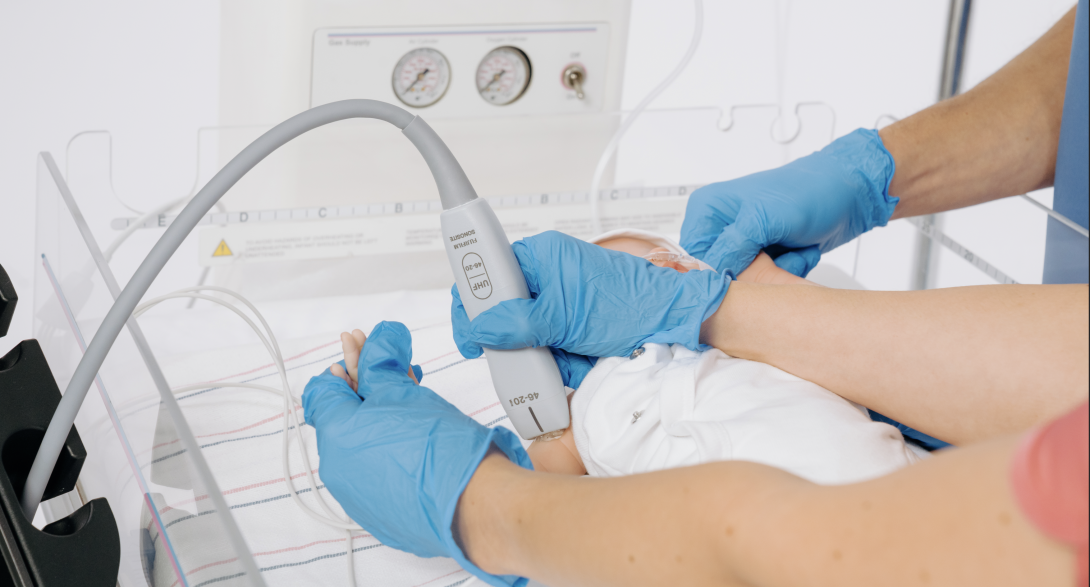
NICU/PICU
Focused care for the smallest patients
Exceptional microanatomy visualization
See superficial submillimeter vessels, nerves, and neonatal anatomy with unmatched clarity in point of care ultrasound, supporting more informed clinical decisions.5,10
Rheumatology
Rheumatology re-imagined
Exceptional microanatomy visualization
See superficial submillimeter anatomy that conventional ultrasound may not capture.1,3,5,6
Aids in diagnosis and intervention
UHF may help you identify subclinical synovitis, erosions, crystal deposits, and inflammation, which may aid you in early diagnosis and intervention in chronic conditions.1,2,3
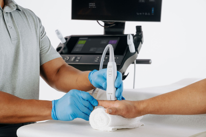
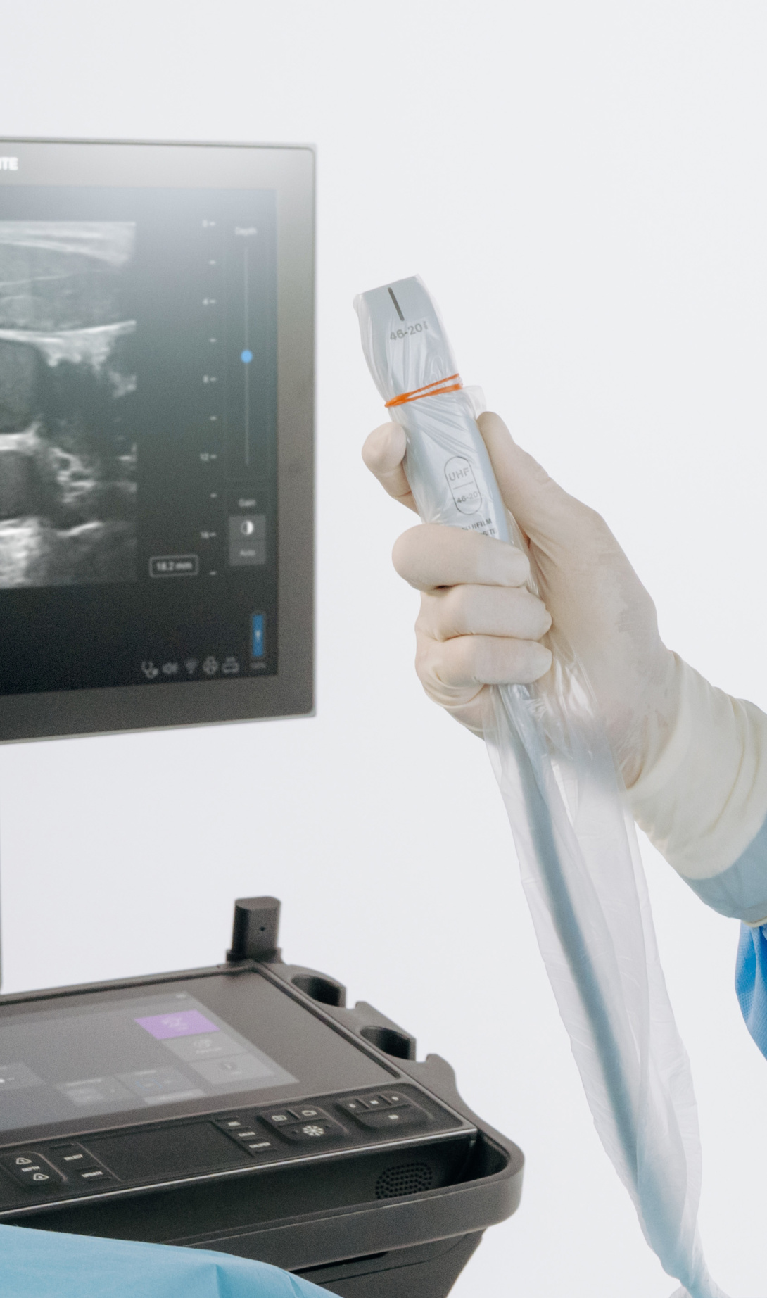
Advanced needle guidance
UHF can provide clear visualization, even in tiny anatomical spaces, to aid in pursuing accuracy and confidence during needle-guided interventions.3,5
Hand surgery
Precision in the palm of your hand
Exceptional microanatomy visualization
See superficial submillimeter anatomy that conventional ultrasound may not capture.1,2,3,6
Confident pre-surgical planning
UHF imaging supports clinicians in mapping anatomy preoperatively.1,2,6
Advanced needle guidance
UHF can provide clear visualization, even in tiny anatomical spaces, to aid in pursuing accuracy and confidence during needle-guided interventions.3,8


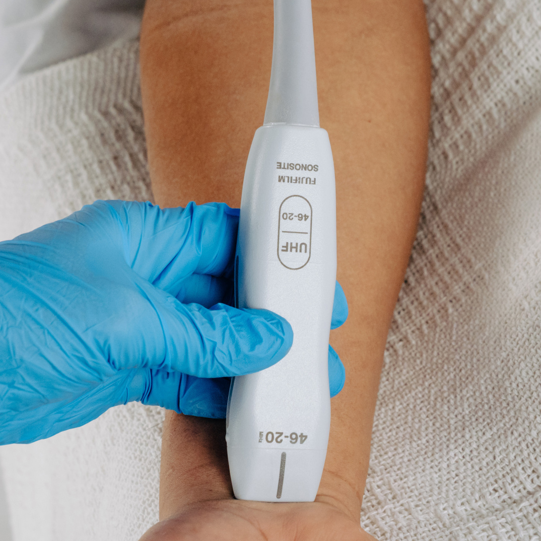
Seeing more matters
As the first and only 46 MHz ultra-high frequency transducer in point-of-care ultrasound, UHF gives you the best resolution for superficial imaging and delivers unmatched clarity within the first centimeter of the imaging field.1,2,3
Microanatomy
See superficial submillimeter anatomy that conventional ultrasound may not capture.1,2
Confidence
UHF can provide clear visualization, even in tiny anatomical spaces, to aid accuracy and confidence during needle-guided interventions.3
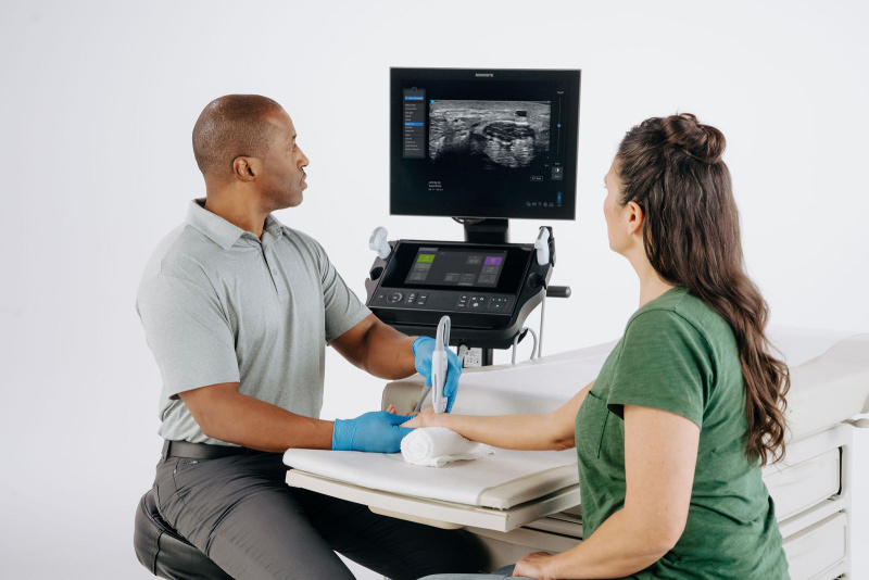
Vascular access
UHF ultrasound may help clinicians improve procedural quality by allowing for improved visualization of superficial anatomical structures.3,4
Improve outcomes, faster
Earlier diagnoses, faster interventions, better care plans—made possible by imaging you can trust.
Take your practice further
Explore what’s possible
46 MHz
The first and only 46 MHz transducer for point-of-care ultrasound.
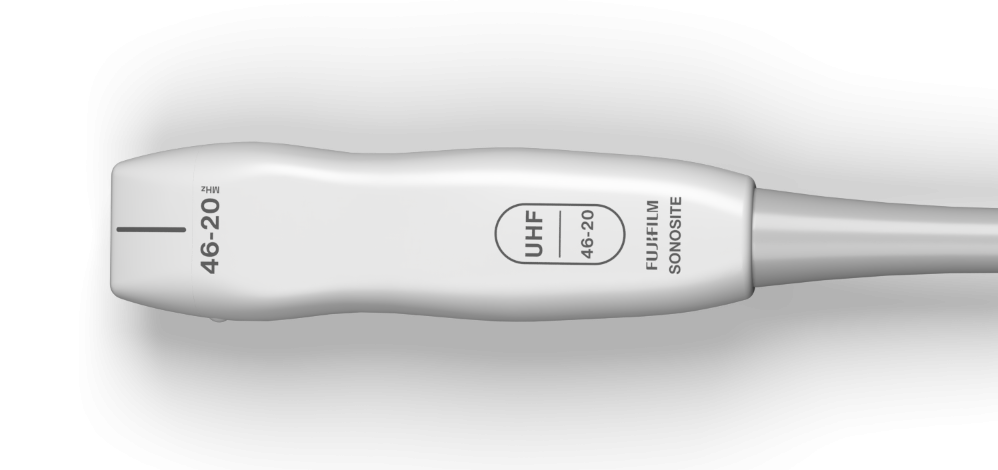
Largest frequency range on any POCUS system.
Research and Publications
Improved GI Diagnostics in Infants Using UHF Ultrasound
Jacobsen RB et al., 2020
This study demonstrates the potential of ultra-high-frequency ultrasound (48–70 MHz) to enhance gastrointestinal diagnostics in infants, providing clearer visualization of bowel wall layers compared to conventional imaging. The results suggest UHF ultrasound may offer valuable support in pediatric GI evaluations.7
Ultrasound in Dermatology: A Comprehensive Review
Almuhanna, N. et al., 2021
This publication reviews current applications of ultrasound in dermatology, including its role in diagnosing inflammatory, neoplastic, and infectious skin diseases. It highlights the advantages of ultrasound in enhancing clinical assessment and guiding treatment decisions.13
UHF Ultrasound in Dermatologic and Aesthetic Medicine
Argalia, G. et al., 2025
This article explores the clinical value of high-frequency and ultra-high-frequency ultrasound in both dermatologic disease management and aesthetic procedures. It discusses imaging benefits for skin lesions, cosmetic filler evaluation, and treatment monitoring.12
Identifying Aganglionosis in Hirschsprung’s Disease with UHF
Hawez, T. et al., 2025
This study evaluates the effectiveness of ultra-high-frequency ultrasound in detecting aganglionosis in Hirschsprung’s disease. The findings support UHF ultrasound as a promising non-invasive adjunct for early diagnosis and surgical planning.11
Clinical Applications of Ultra-High-Frequency Ultrasound
Russo, A. et al., 2022
This comprehensive review outlines a wide range of clinical applications for ultra-high-frequency ultrasound across multiple specialties, from dermatology to musculoskeletal imaging. The article emphasizes its precision in superficial tissue evaluation.2
Imaging Hyaluronic Acid Fillers with UHF Ultrasound
Salvia, G. et al., 2023
This research highlights the use of ultra-high-frequency ultrasound for visualizing hyaluronic acid fillers in the nasolabial folds, offering detailed assessment of filler placement and integrity. It supports the use of UHF imaging in aesthetic medicine for pre- and post-treatment evaluation.10
UHF Ultrasound as a Diagnostic Tool in Hand Surgery
Viviano, S. L. et al., 2018
This study explores the utility of ultra-high frequency ultrasound in diagnosing hand pathologies relevant to surgical planning. The authors highlight its ability to provide detailed imaging of tendons, nerves, and soft tissue structures, enhancing diagnostic accuracy in hand surgery.6
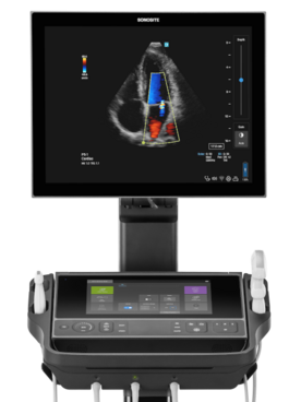
Sonosite LX
Unmatched frequency range
Sonosite LX has the largest frequency range on any POCUS system, making it the most comprehensive imaging solution on the market today.
- Albano, D., Aringhieri, G., Messina, C., De Flaviis, L., & Sconfienza, L. M. (2020). High-Frequency and Ultra-High Frequency Ultrasound: Musculoskeletal Imaging up to 70 MHz. Seminars in musculoskeletal radiology, 24(2), 125–134. https://doi.org/10.1055/s-0039-3401042
- Russo, A.; Reginelli, A.; Lacasella, G.V.; Grassi, E.; Karaboue, M.A.A.; Quarto, T.; Busetto, G.M.; Aliprandi, A.; Grassi, R.; Berritto, D. Clinical Application of Ultra-HighFrequency Ultrasound. J. Pers. Med. 2022, 12, 1733. https://doi.org/10.3390/jpm12101733
- Ait Ichou, J., Gauvin, S., & Faingold, R. (2021). Ultra-high-frequency ultrasound of superficial and musculoskeletal structures in the pediatric population. Pediatric radiology, 51(9), 1748–1757. https://doi.org/10.1007/s00247-021-04978-0
- Hayashi, A., Giacalone, G., Yamamoto, T., Belva, F., Visconti, G., Hayashi, N., Handa, M., Yoshimatsu, H., & Salgarello, M. (2019). Ultra High-frequency Ultrasonographic Imaging with 70 MHz Scanner for Visualization of the Lymphatic Vessels. Plastic and reconstructive surgery. Global open, 7(1), e2086. https://doi.org/10.1097/GOX.0000000000002086
- Latham, G. J., Veneracion, M. L., Joffe, D. C., Bosenberg, A. T., Flack, S. H., & Low, D. K. (2013). High-frequency micro-ultrasound for vascular access in young children--a feasibility study by the High-frequency UltraSound in Kids studY (HUSKY) group. Paediatric anaesthesia, 23(6), 529–535. https://doi.org/10.1111/pan.12131
- Viviano, S. L., Chandler, L. K., & Keith, J. D. (2018). Ultrahigh Frequency Ultrasound Imaging of the Hand: A New Diagnostic Tool for Hand Surgery. Hand (New York, N.Y.), 13(6), 720–725.https://doi.org/10.1177/1558944717731856
- Jacobsen RB, Hebelka H, Gatzinsky V, Elfvin A, Dangardt F. Ultra-high-frequency ultrasound (48–70 MHz) is a promising tool for improved gastrointestinal diagnostics in infants. Acta Paediatr. 2024; 113: 2304–2311. https://doi.org/10.1111/apa.17342
- Brusciano, V., & Lecce, M. (2024). Advantages of the use of ultrasound in newborn vascular access: a systematic review. Journal of ultrasound, 27(2), 203–207. https://doi.org/10.1007/s40477-023-00832-1
- Currie M, Vashisht R, Elkin D, et al. Ultrasound Intravascular Access. [Updated 2024 Jul 2]. In: StatPearls [Internet]. Treasure Island (FL): StatPearls Publishing; 2025 Jan-. Available from:https://www.ncbi.nlm.nih.gov/books/NBK448093/
- Salvia, G.; Zerbinati, N.; Manzo Margiotta, F.; Michelucci, A.; Granieri, G.; Fidanzi, C.; Morganti, R.; Romanelli, M.; Dini, V. Ultra-High-Frequency Ultrasound as an Innovative Imaging Evaluation of Hyaluronic Acid Filler in Nasolabial Folds. Diagnostics 2023, 13, 2761. https://doi.org/10.3390/ diagnostics13172761
- Hawez, T., Evertsson, M., Erlöv, T. et al. The use of ultra-high frequency ultrasound in identifying aganglionosis in Hirschsprung’s disease. Sci Rep 15, 15124 (2025). https://doi.org/10.1038/s41598-025-99897-7
- Argalia, G.; Reginelli, A.; Molinelli, E.; Russo, A.; Michelucci, A.; Sechi, A.; Marzano, A.V.; Desyatnikova, S.; Fogante, M.; Patanè, V.; et al. High-Frequency and Ultra-High-Frequency Ultrasound in Dermatologic Diseases and Aesthetic Medicine. Medicina 2025, 61, 220. https://doi.org/10.3390/medicina61020220
- Almuhanna, N., Wortsman, X., Wohlmuth-Wieser, I., Kinoshita-Ise, M., & Alhusayen, R. (2021). Overview of Ultrasound Imaging Applications in Dermatology. Journal of cutaneous medicine and surgery, 25(5), 521–529. https://doi.org/10.1177/1203475421999326