


Transcript
- So welcome to the Sonosite Behind the Scan webinar,
titled "TEU in the ICU: A Multi-tool for the Intensivist".
My name is Chris Pennell,
and I'll be hosting today's webinar.
Before we begin, please be advised all attendees are muted.
As the discussion is going,
you can type your questions into the Q&A box at any time
in the toolbar located at either the bottom
or the side of your screen,
and we'll get to those questions at the Q&A session
at the end of the discussion.
This webinar will be recorded
and archived for future reference.
And another thing for today's presentation,
today's presentation includes
an interactive polling element.
So to participate in that,
you can text NV303 to 22333.
And then when a poll appears on the screen,
you can text your answer to that number
and see it reflected on the poll.
So here with us today, we have Dr. Nick Villalobos.
Dr. Villalobos is the medical intensive care unit director
at the Brooke Army Medical Center
and assistant professor of medicine
for the Uniformed Services University in San Antonio, Texas.
He completed his training at the University of New Mexico
and holds a board certification
in critical care echocardiography.
He's part of a small group of physicians
for the critical care air transport team
in the United States Air Force
and has deployed with the military's ECMO transport team.
Dr. Villalobos is an ICU medical director
and the director of ultrasonography
for the fellowship and residency program
at the Brooke Army Medical Center.
And with that, I'll turn it over to Dr. Villalobos
to get started.
- Thanks for the intro, Chris,
and thanks for everybody that logged on.
Hopefully, this is useful to everyone.
You know, it's a interesting topic
and it's kind of gaining some steam in the ICU.
So I have about 40 minutes
to try to empower everyone here on the Zoom call
that transesophageal ultrasound can be used by intensivists.
So I'll start off with my disclosure.
I am an active duty service member.
First and foremost, I want to thank everybody
that I am able to work with for the dedication
and the willingness to sacrifice themselves for our country.
So that's my one disclosure.
The goals today are set forth here.
I'm pretty sure everybody could read it.
Again, I have about 30 to 40 minutes
to really kind of empower everybody
on transesophageal ultrasound in the ICU.
All right, so we'll start off with the poll.
I'll give you guys a couple minutes to text the number above
and A, B, C, or D,
and then we'll see kind of
where everybody's background is coming from.
And again, I'll point out that if you guys have questions
during the presentation, go ahead and type 'em
in the chat or virtually raise your hand
and I'll try to get to those after the presentation is done.
Okay.
All right, so seems like some people are working
on training, some people just want to learn more,
which is great.
All right, so we'll move forward.
All right, so I'll start with my inclusion criteria,
and this is supported
by the 2013 American Society of Echocardiography.
So really low threshold to drop a transesophageal probe.
So A, I'm not getting good enough transthoracic views.
B, do they have an esophagus?
And C, is the airway protected?
So duck, duck, goose.
This is the prototypical image
that you guys see at the bottom
that elicits my response
in placing a transesophageal probe to get a better view.
And much like I think most of the people,
if not everybody here on the webinar,
were using point-of-care ultrasound every day
in our practice.
So what better reason
than to get more impactful views do we need
to drop a transesophageal probe?
And some people have seen this picture before.
This is a cardiologist
and an anesthesiologist protecting their playground.
I'm joking.
You know, cardiologists
and anesthesiologists have definitely been at the forefront
of using transesophageal ultrasound.
I think I do get a lot of eyebrows raised initially
when I started doing transesophageal ultrasound in the ICU.
But when you use it in a focused approach in the ICU,
the cardiologist and the anesthesiology group
will applaud you for focusing and helping
to find the answers that your patients need.
All right, so it's a multi-tool.
Why do I call it a multi-tool?
We're making countless predictions every day in the ICU.
Using transesophageal ultrasound is going to help you
with your decision-making in making those predictions.
It's also useful whenever you're doing procedures.
So you're not only helping the decision-making,
you're actually helping yourself with those procedures.
In my practice, I'm cannulating ECMO or decannulating ECMO
or even following a patient on ECMO
throughout their hospital course.
Transesophageal ultrasound helps me
make those daily decisions on these patients.
So usually what I ask my trainees to do
before they start implementing this into their practice is
to get to a point where they feel comfortable enough
where they can take the critical care echo exam.
I don't necessarily ask anybody that they have
to take the boards,
but you do need a fundamental background in ultrasonography
and echocardiography to really implement the data
that you get from transesophageal ultrasound.
So the boards were implemented back in 2019,
so a pretty new thing for intensivists.
They also include surface,
so transthoracic studies
and transesophageal echocardiography.
For the intensivists, really,
you only need to get six views versus the 20 views
that the cardiologists are getting for the ASE.
The other thing I'm going to point out to you guys
during the presentation is the comparison,
and really just to contrast between pulmonary artery cath.
I don't want you to necessarily think
that this is a head-to-head comparison
between transesophageal ultrasound and Swans.
More just a different modality
that you can potentially use in the ICU,
again, to help generate that data that you need
to make a decision.
And the other question that I get oftentimes is,
"Do you think that you're as good as a cardiologist?"
And the answer is no.
I will never be at that level,
nor is my practice focused on that specifically.
In the ICU, we have a list of questions
that we're trying to answer,
and if I focus my examination on those questions,
then I'm able to generate some information to, again,
get towards that answer that I'm looking for.
So there's a lot of research out there
that shows that whenever we focus our study,
we can actually generate that useful information
that can be interpreted as well as a cardiologist.
And again, background, I'm a pulmonologist.
So I think, you know,
I think I was asked to give this presentation more
so to demonstrate to the people listening
that if me, a simple Texan pulmonologist,
can figure this out and use this in their everyday practice,
I'm sure anybody on this webinar can.
So a couple points made from a few of the studies
that you see below, all fairly new studies,
kind of reading between the lines.
This is a fairly low-risk procedure that you're doing,
and it's actually pretty impactful.
I mean if you're able to couple this with your examination
that you can't quite get that view that you want,
you could really turn the prognosis around
in a certain patient population.
So I gave you guys my inclusion criteria.
This is more so what you think a potential contraindication
for transesophageal ultrasound is.
So again, I mentioned it's a fairly low-risk procedure.
Sorry, you couldn't read all of those but...
So, I mean, do you think,
you know, having too many pressors on, low platelets,
maybe having a difficult airway,
or maybe being too obese is a contraindication?
I'll kind of let you guys know
what my contraindications are.
So yeah, I agree with everybody. Really, none of these are.
I mean like I mentioned in the previous slide,
they've done studies even on patients
that have a low fibrinogen level,
elevated INR, low platelets,
they could be on a number of pressors,
and really, that's the reason why we want to do this study
to write, to try to get down to the nuts and bolts
of the patient's physiology.
Really, if they don't have an esophagus
or if they're profusely bleeding from their esophagus,
I probably won't drop the probe, but aside from that,
few and far between contraindications for this.
All right.
So I'll try to run through a couple cases for you guys
to show how there are different ways
that I'm using transesophageal ultrasound
in one particular setting or one patient
to help drive the decisions being made.
So this is a 30-year-old female,
gunshot wound of the chest
that recently had a thoracotomy done for RV injury.
The bullet was removed
and ended up bouncing around in the thorax.
Patient got a gastrectomy.
I get a call from my fellow, you know, they're saying,
"Hey, not sure if she's still bleeding.
We don't really know what's going on.
She's on three pressers now.
Do you mind dropping the probe?"
"So yep, I'll be there in no time."
So I get my trusty rusty M-Turbo,
and yes, this does do transesophageal ultrasound.
This is an old-school machine
and that's why I love it though.
It's built for tough as they say.
So we go to the patient,
we drop the probe right in time for us to do a bubble study.
So I'll start off kind of going
through the fundamental views for transesophageal ultrasound
and that's starting off
with the mid-esophageal four chamber.
So you see, we're doing a bubble study here,
making sure we don't have an ASD or a VSD.
This is essentially a four-chamber
apical four view on surface.
And like I mentioned, you guys,
we're going to kind of compare and contrast
between transesophageal ultrasound surface
and pulmonary artery cath between what we're able to get.
So really, this is our home row.
We're dropping the probe 40 to 45 centimeters
into the esophagus.
We're omniplaning at zero.
So we're not really looking around using
that easy button to omniplane
to get a nice home row fundamental view.
So for this patient, we did a bubble study,
didn't see anything significant kind of qualitatively.
I noticed that the LV's potentially a little dilated,
maybe not squeezing as much as I wanted to.
After the bubble study,
kind of looking around a little bit more,
that RV to LV ratio is potentially one-to-one.
I see some interventricular interdependence,
you kind of see that septum bouncing around.
So we're gathering useful information.
I'm not going to make a strong prediction at this point,
probably going to want to look a little bit more.
So then we move on to our mid-esophageal long
and our two-chamber view.
So this is omniplaning from 90 to 120 degrees
to get that mid-esophageal long axis view.
And in this view,
we can get a lot more information
regarding the aortic valve,
a little bit more about our cardiac
or ejection fraction more so than the output at this point.
We're able to rule out dynamic LVOT obstruction
or rule it in,
and we are able to get an idea about our filling pressures.
You kind of see that I have a plus or minus both on surface
and transesophageal echo.
No, I can't tell you what the pressures are like a Swan can,
but I can get an idea based off of the size of the chambers.
So in this case,
I'm noticing that the LV is actually maybe a little bit more
ballooned out than I was expecting
on that four-chamber view.
And this is the right ventricle,
for some of you guys that haven't seen this view before,
kind of going through it, the left atrium, left ventricle,
aorta, and the right ventricle.
Right ventricle here looks a little large,
kind of how we were guesstimating
on that four-chamber view, potentially overloaded.
Here's another clip of a patient
that has a dynamic LVOT obstruction.
Up to a quarter of our patients coming in with septic shock
into the ICU will have upfront a dynamic LVOT obstruction.
So this is a great tool to use
whenever you get a distributive shock patient
that you're kind of lost at what to do next.
Here's another clip where this is
in between ACLS.
We have a pulse check going on.
This is what you think is PEA arrest.
At this point, I would shock this patient
out of their coarse V-fib.
Here's another view with an Impella.
So monitoring your devices
using transesophageal ultrasound is extremely easy compared
to a difficult body habitus patient or someone
with very elevated ventilator settings
whenever you have an Impella or an LVAD or ECMO.
So moving on to our mid-esophageal bicaval view.
So at this point,
we decided to put this patient on VA ECMO for BiV failure.
Raise your hand if you've ever heard of or seen a patient
that you're placing onto ECMO get an RV perforation
from the wires.
So the last thing that you want to do is have
that stiff Amplatz wire go straight
into the RV and cause a perforation.
So using transesophageal ultrasound,
you'll be able to actually watch the wire go from,
for a drainage cannula,
watch the wire go from the IVC to the SVC to make sure
that you're appropriately placing that ECMO cannula.
So with the bicaval view,
you're also able to see the SVC more clearly
where it's really difficult.
I know there's some people that are doing it
out there on surface,
but again, this is a more accurate measurement
of your volume responsiveness using the SVC.
Again, doing that bicaval view is really going to help out
with your procedures, whether you're cannulating for ECMO
or placing a transvenous pacer
or even a Swan, for that matter.
And you're actually able to see, so here's your IVC,
your SVC and the entrance into the right atrium.
Here's your left atrium.
You can actually diagnose shunts
that way using the mid-esophageal bicaval view.
Here's another patient.
This is a gentleman that we had in the burn unit
that had 40% body surface area burns to his lower extremity.
So we ended up picking a Crescent dual-lumen ECMO cannula.
If you guys have had any experience with those,
placement is often difficult
because the inflow port is
in between the drainage cannulas.
So using transesophageal echo,
we were actually able to place the inflow port exactly
where we wanted to to have the jet directed
into the right atrium, and that's just with color doppler.
I know some people will inject bubbles,
and that can be used.
And you can also watch your cannula go from the SVC
into the IVC in real time to make sure you're going
where you want to with your cannula.
So really key four procedures in that way.
This is back to our 30-year-old gunshot wound.
Whenever you're cannulating ECMO,
whether it be in ECPR or in kind of a,
you know, all-hands-on-deck situation,
you want to actually make sure you're cannulating VA ECMO
and not VV.
So confirming that your wires in the aorta,
and in this case,
you could see our wire going into the aortic arch.
So we're confirming wire placement with our aortic view.
And you know, really useful.
This is a patient after chest compressions.
During transesophageal echo,
we're actually able to diagnose a dissection,
and this is with color through that dissection.
So really, really useful,
especially after an arrest or a peri-arrest.
So going back to our gunshot wound patient.
So this is a clip after we've placed the patient on ECMO,
we're kind of noticing now that, all right,
this patient is probably volume down at this point.
We're able to kind of get an idea
of what the LV pressures are,
and volume, as you can see significantly decreased.
Start looking around a little bit more
and we see something new here.
We see that this patient is coagulopathic,
probably from their recent trauma.
And during ECMO cannulation,
we've actually introduced a little thrombus into the RV.
In this case, it's VA ECMO, so we're not as concerned,
but again, if you're cannulating for VV ECMO
and you get a large clot,
the risk is that clot going back into the oxygenator
and you having potentially failure of your ECMO circuit.
So periprocedural monitoring is paramount
and another significant use that you get
from the transesophageal ultrasound.
And in this case,
we go back to our mid-esophageal long axis view
and look again.
So let's say that, you know,
and that's what this case was, we're late in the evening,
we're still in the midst of our resuscitation
in terms of blood product from this traumatic patient,
placed the patient on VA ECMO.
This may and this did kind of elicit us
to talk to cardiology about putting an LV vent,
so an Impella,
noticing that the LV was starting to enlarge
and the RV was still enlarged.
So kind of the, again,
another point for the peri-procedure monitoring.
All right, this is a prototypical case that I'm sure many
of you working in the cardiac ICU have seen.
Whoops, got dark on me.
We got a 54-year-old patient that recently had a CABG.
In the OR, anesthesia is
at least saying things were pretty good other
than getting a little methylene blue
and coming out on a, quote, unquote, "whiff of pressor."
So you know, within an hour or two,
this patient had an increasing pressor requirement.
Of course, we don't get really good surface view,
so we drop the probe,
and this is our transgastric short axis view.
So what we're able to see here,
where I think it drives a really good point
for us using transesophageal ultrasound is wall motion.
So this patient recently had a CABG.
This will prompt me to ask the surgeon,
"Hey, do you think that graph
that you just placed is working like it's supposed to?"
Here's another clip of a transgastric view,
and this is a transgastric deep where you guys see
on this chart, I have cardiac output.
So as you guys are probably already using
your surface echo for,
is getting a VTI of that LVOT.
This is essentially the same image
that you'll get on an apical five-chamber view
to get your VTI to estimate the cardiac output.
This is another patient that has a,
you know, the prototypical D sign
and a very underfilled left ventricle,
again with that transgastric view.
So going back to this case number two,
our post-CABG patient,
we're looking around a little bit more.
We're seeing that the LV function doesn't look so good,
and then I get another inkling
about what may potentially be going on in this case.
So kind of looking around here,
I see something on the outside
of the myocardium, pericardium.
This is a little modified view
that we won't go over in this webinar,
but again, useful.
There's a Swan in place.
We could kind of see that the RV, which is right here,
is not squeezing as much as we would want it to.
And what we notice is that there's a significant clot burden
around the RV.
And you also notice
that the right atrium is bulging into the left atrium.
So seems like there's elevated RA pressures.
So this patient ended up having a graft that was down.
So that transgastric view got us speaking
with the cardiothoracic surgeon
and asking them to go to the cath lab
to verify that the graft was functional,
which it wasn't at that point.
Okay, a little bit on hemodynamics.
This is a clip from that case one that we had,
that gunshot wound.
We put color through the tricuspid valve
and noticed that there was significant tricuspid regurge,
signifying that there's probably a certain degree
of elevated pulmonary artery systolic pressure.
Another use that I mentioned to you guys earlier is looking
at that SVC.
There has been evidence that shows
that you have a little bit more accuracy
in determining volume responsiveness,
looking at the SVC, which is intrathoracic,
versus your IVC, which is in the abdomen.
Another thing that I love
to use the transesophageal ultrasound
for is titrating the ventilator.
This is a recent patient that we had on VV ECMO.
A lot of us are used to putting the patient on,
quote, unquote "rest settings."
There's been some recent evidence that shows that
that approach may not work for everybody.
So this patient had,
in the aorta view, had consolidation of their lung.
You can see the static air bronchograms,
and then after a little PEEP titration,
so what we did is titrate the PEEP up
by two and then we would go back and look at the RV
to make sure that we weren't falling behind
with our RV overload,
and we were able to recruit that lung.
Within the course of five minutes,
we're able to get a response and go from consolidation
and air bronchograms to the development of the B-lines.
So we're able to recruit the lung,
not fall back on our RV
or develop RV failure while we're doing that
and also maintain an adequate driving pressure.
I mentioned efficiency and practice.
You have to keep in mind
whenever you're doing other procedures,
how long is it going to take to do certain things.
I know for me,
I'm not the the fastest at dropping in a Swan,
but when it comes to transesophageal ultrasound,
there have been studies that show that a provider
with minimal experience can get a complete exam
from placing the probe in to completing the exam
in less than 20 minutes.
So also looking at your day, if you have a busy ICU,
how much time do you have to devote
to doing a comprehensive exam?
This garners a lot of its utility.
So this is not a statistics talk,
but if you guys have heard
of the Bayesian model of statistics, in my interpretation,
it's kind of reading in between the lines
of a lot of the studies that are out there.
So I like to apply that to transesophageal ultrasound.
I'm using POCUS every day in my practice,
and this may not in and itself
on a large randomized control trial prove
that there is a mortality benefit, but in my practice,
I find a lot of benefit
from using transesophageal ultrasound.
So this is a good panel that I stole
from one of my colleagues showing ECPR in progress.
So this is chest compressions on the top left.
You see nice recoil and filling of the left ventricle.
Looks like chest compressions are very adequate.
This is a pulse check
where you see essentially cardiac standstill.
This, on the bottom left,
is introduction of that arterial limb
of the ECMO cannula wire going into the descending aorta.
And this is afterwards,
after we've put the patient on VA ECMO.
So really, really neat
and quick immediate information.
Again, this is going back to our patient
that has information that keeps on giving.
After we placed the patient on VA ECMO,
we were worried that we were having bleeding from somewhere.
So this is a long axis of that descending aorta.
And what we're seeing here, we see lung,
and this is actually a large hemothorax.
So this patient had chest tubes in place,
but during the cannulation,
seems like there was a clot
that developed in one of the chest tubes.
So I asked the resident, "Hey, milk that chest tube,
and try to get that clot out of there."
Again, we get immediate feedback.
So within seconds, that hemothorax starts to clear up
and we're seeing more of that lung parenchyma
after they get that clot out of the chest tube.
This is a different patient,
one that came
in shock with hypoxia.
This is an upper esophageal pulmonary artery view.
This is the right pulmonary artery.
We see a little hyperechoic density here in the right PA.
To confirm our suspicion, we put color through that area,
and we've diagnosed a massive pulmonary embolism
and we did that before we had to go to the CT scanner.
So last question I have for you guys,
and I get this question asked a lot,
how long do you keep the probe in
and when do you take it out?
I'll give you guys a minute or two to submit your answers.
Let's see how we're doing.
Yeah, so usually the cardiologist will remove the probe
right after the exam.
But us, like I mentioned,
transesophageal ultrasound derives a lot
of its importance in the ICU
for the ability for the intensivist
to be able to trend our decisions that we make.
So in my practice, as best as possible,
I will keep the probe in, potentially the entire shift.
As long as I'm actually using that data
that I gather from the transesophageal ultrasound
and it's useful to me,
I will keep it in as long as I need to.
I personally don't leave it in 24 hours.
This may be a question to some other folks
that use it regularly as well.
But in my practice,
I'll leave it in potentially an hour or two hours,
three hours or an entire shift.
And again, as long as the data is useful,
then you have all the backing to do that.
So a quick little thing on credentialing and programming,
again, the way that I usually ask my trainees
to approach this is if they've gathered enough expertise
in transthoracic echocardiogram and feel comfortable taking
that boards to even get the (indistinct) status.
All I ask them to do is 10 studies,
10 transesophageal ultrasound studies with me
to independently practice.
Some programs or some people ask their trainees
to do upwards of 50 studies.
If you are out in practice alone
and you're trying to implement this into your practice,
I would probably lean more towards trying to get,
you know, anywhere between 20 to 50 studies
with an anesthesiologist or with a cardiologist.
But if you're in a training program that already offers
intensivist-driven transesophageal ultrasound,
that threshold could be potentially lowered.
The other thing that you absolutely need to do,
especially if you're billing for this study,
is you need to have a repository
for those clips that you're getting,
and you need to be able to quality review them
with somebody that is either board certified
or has expertise doing transesophageal ultrasound.
This is the CPT code if you're going to start billing
for transesophageal ultrasound.
You need to place in a procedure note
and you need to have a description of the procedure
with your assessment.
And usually, it's fairly easy in a place that has an OR
because there's probably anesthesiologist
or a cardiologist already doing this,
but you need to have biomed approval
with the appropriate cleaning strategy.
So this is the Medicare-Medicaid
compensation for point-of-care transesophageal ultrasound.
And what I have in the box is between the physician
and the facility, this is what you're getting reimbursed.
So no, this isn't a very lucrative procedure
that you're doing, but again,
that's not why we're doing this.
So I will do a transesophageal ultrasound
probably a couple times a week.
So really if you're doing this regularly,
which you should if you consider adding this
into your practice, it pays for itself pretty quickly.
And that's all I have for you guys.
That's my email, so please feel free to reach out to me
if you have any questions on how to introduce this
into your practice, on why you would do this,
on anything interesting clips-wise, cases-wise.
I'm also on LinkedIn. I'm not a big Twitter person.
Below, I have a really great online source
for anybody getting into it.
All you have to do is Google
the Virtual Transesophageal Echocardiography program
at Toronto, and they have nice virtual clips
for you to go through.
And also big thanks to Dr. Wray
at the University of New Mexico for letting me steal some
of his clips.
All right.
- [Chris] All right, well, thanks, Dr. Villalobos,
for your awesome presentation.
You really covered a lot of stuff in this.
So it is time for questions.
So if anybody does have any questions,
feel free to put them in the Q&A box either at the bottom
or the side of your screen.
We do have a few already in here.
"How do we obtain the 150 TEE for certification?"
- So I'm glad you brought that question up.
So that's kind
of specifically towards the board certification
for critical care echocardiography,
and it's 150 transthoracic views.
I kind of put that up there as a marker
if somebody wants to get that board certification
through the National Board of Echocardiography.
Really, if you're trying to get studies under your belt
and you're at a place
that doesn't have intensivists already doing this,
I would reach out to either anesthesia or cardiology
to get some practice under them,
and they're usually receptive if you mention to them,
"Hey, I'm an intensivist, I just want to do a focused exam.
I'm looking to just gather six views,
really looking at that four-chamber view,
the long axis view, and a transgastric view,
and maybe a bicaval."
I haven't had any problems with any anesthesiologists
or cardiologists kind of taking people under their wing
and introducing them that way.
Or there are, you know,
I will say that there are programs like CHEST or SCCM
that are offering programs
for kind of an introduction into transesophageal ultrasound.
- [Chris] All right.
We also have,
"Do you need a like different ultrasound system
to perform a TEU?"
- No, most systems that you probably already have,
like I showed you guys, my M-Turbo,
you could hook up a probe to it,
and I'm sure everybody from Sonosite
on this call will also echo that,
that every single one of their machines is TEU-compatible.
- [Chris] Excellent, and, "Do you have any idea
how many like hospitals or programs are like getting ready
to like implement TEU in their practices?"
- That's a really good question.
I will say that in the ICU, it's a relatively,
I wouldn't say nascent thing, but in terms of its broad use,
it's pretty nascent.
You know, I can say that I came from a facility
that had its regular use and we were doing ECPR
and we garnered a lot of expertise from that program,
and it's kind of been spreading out throughout the country.
But you know, the literature's been out
since the early 2000s,
and intensivists using it recently reached out
to one of the, you know, quote, unquote,
"godfathers of transesophageal ultrasound,"
here in the United States, and he kind of told me,
he said, you know,
"America is about 10 years behind Europe
in its implementation, but I think, you know,
I think it's going to be become common practice."
I know the emergency department has already put statements
out through ACEP on its expected use
for emergency medicine docs.
And I think in the ICU,
it's only a matter of time
before it becomes regularly implemented.
- [Chris] Excellent.
All right, well,
it doesn't look like we have any more questions.
So Dr. Villalobos, thank you so much for coming here
and presenting about this.
Like I said, it was a really excellent presentation.
And yeah, if anybody has any questions after the fact,
as Dr. Villalobos said, you can go ahead and email him.
And thank you all very much for coming
and we'll see you at the next behind the set,
(laughs) Behind the Scan Sonosite webinar.
Bye-bye.
Transesophageal Ultrasound (TEU) has become essential in Critical Care settings, offering point-of-care clinicians a valuable tool for cardiac and hemodynamic status assessment and during resuscitation.
What You'll Learn
Upon viewing this webinar, you will be able to provide better patient care by having a greater understanding of:
- Patient selection and limitations for TEU in the ICU.
- Transesophageal Ultrasound views for typical ICU use, and when each view is appropriate.
- Key hemodynamic principles for critical care echocardiography and rules to abide by in ECMO cannulation, monitoring, and weaning.
- Credentialing and programming for intensivists.
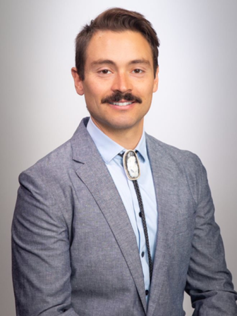
Dr. Nick Villalobos is an Assistant Professor of Medicine for the Uniformed Services University at the San Antonio Military Medical Center in the Division of Pulmonary, Critical Care, and ECMO. He completed his training at the University of New Mexico and holds a board certification in Critical Care Echocardiography. He is part of a small group of physicians for the critical care air transport team in the United States Air Force and has deployed with the military's ECMO transport team. Dr. Villalobos is an ICU medical director and the director of ultrasonography for the fellowship and residency program.


This educational webinar is intended for healthcare professionals and not for patients or consumers. The material is provided for general educational purposes, as a reference and a supplement to professional experience, education and training, and should not be considered the exclusive source for this type of information. The presenter in this webinar is a paid consultant of FUJIFILM SonoSite, Inc., and is sharing the presenter's own views and opinions that do not necessarily represent the views and opinions of FUJIFILM SonoSite, Inc. This educational webinar is not intended to recommend any device for a particular indication or to provide indications for use for any device. At all times, it is the professional responsibility of the practitioner to exercise independent clinical judgment in each particular situation. Fujifilm assumes no responsibility or liability for any misuse of the information imparted in this webinar. This educational webinar does not supplement, replace, or supersede device labeling, including instructions for use, which accompanies any FUJIFILM Sonosite product.