


Transcript
- Welcome to the Sono Site webinar today titled Point
of Care, echocardiography and Critical Care.
It's my pleasure to introduce our speaker today. Dr.
Seth Konig is the Chief of Medicine, chief
of Pulmonary Critical Care
and Sleep Medicine at Kent Hospital in Rhode Island.
Dr. Konig has been interested in bringing point
of care ultrasound to critical care for over a decade.
His efforts and those of his associates
and colleagues have helped
revolutionize the way critically ill.
Patients are managed in the US and around the globe. Dr.
Koenig's research interests have led to numerous studies
and publications involving point of care, ultra sonography,
particularly in critical care.
And thank you so much for being here today, Dr. Koenig.
And with that, I will turn it over to you.
- Thank you. With all that talk, actually,
I'm just a doctor who loves to take care of patients
and I think that that's a really important
part of what we do.
Nothing makes me happier than being a soldier next
to the battlefield of medicine.
And all I wanna do over the next 20,
30 minutes is just share some of the ideas over the years
that we've built up on focusing in on critically ill
patients when we need information quickly,
how do we get it in the most efficient manner,
in the most accurate manner?
And probably if you're on this, this webinar, this new world
of ours, the zoom world,
you are probably already have drank the Kool-Aid
and hopefully are beginning to use it
or have become quite accustomed to it.
And so I think I titled this, our patients deserve it
because just like everything else in medicine, we wouldn't,
we, we, we always try to first use our history
and physical in our brains and then we go ahead
and make up a differential diagnosis and then we go ahead
and use our technology to either confirm
or deny what we think.
So I'm just gonna go through very simply some
of these objectives, which, you know,
most people are starting to understand maybe
what constitutes point of care echocardiography,
but it's important obviously for all of us to understand
how it differs from our cardiology colleagues when they ask
them to do an echocardiography.
I think it's important to familiarize oneself
with the basic echocardiographic views.
And this way we speak the same language when we're
talking about things.
I think it's important to know what some
of the resources are that are available.
And, and again, this will just be my opinion about
what is necessary sort of to become competent in point
of care echocardiography, because resources are one thing,
but the personal sweat
that we put into learning these things is really
what makes folks become competent.
And then I have a couple of cases I think
that may drive some of these points home in regards
to how do we actually do it at the bedside and,
and what is it that we're trying to accomplish?
So what is a pocus echocardiogram versus standard
cardiology driven exam?
And I think maybe there are three points is
there could be more.
People could probably come up with different things,
but the main thing is
that the clinical provider is the person who both
or who performs, then interprets
and then clinically integrates the findings right then
and there for that patient.
And that means there's no clinical or time dissociation.
And that's an important observation,
meaning there's no clinical dissociation.
That is, you're not asking somebody else
to do an examination, which is then read by a third party
who is not really invested in the, the, the patient.
Nothing wrong with that, it's just that they're not there,
it's offline.
And then of course, time dissociation is the fact
that you may want an echo right this the second.
It could take an hour or two to get if you're lucky.
But then also there's that, that lack of time
or that time when someone has to read it.
And so that's the first thing.
And the second thing, there's no clinical
and time dissociation and the clinical provider performs
and interprets and integrates the findings.
But also a main thing is you're trying
to answer an urgent question, why is my patient in shock?
Why is my patient in respiratory failure?
And so all you're, what you're trying
to do is use a minimal number of views
to answer a clinical question.
And it's sort of the basic focus.
Echocardiogram is started with no color or spectral doppler.
And now some, some people feel mini, you know, there,
there there's a minimum amount that you can do
that answers a lot of questions.
And we, I can share some of that with,
so the cardiology exam, just so everybody's aware
of the difference
and one of the main complaints that used to come out
of cardiology, which is no longer now cardiology
recognizes keenly the importance of of,
of appropriate pocus echocardiogram.
And what I mean by that is folks
who have become competent at performing it
and know when they don't know
and when it's time to ask for advice.
And so a cardiology exam,
remember it's done exactly the same way every time.
It's a full examination with lots of views, full doppler,
both color and spectral with chamber quantification.
Very important stuff.
But if you're trying to figure out if a patient is in shock
from a massive pe, it might be that you only need one view,
one view that just shows a dilated not functioning
very well, right?
Ventricle may be all you need,
especially if you add in a DVT study and find A DVT.
And that's the difference you're asking
and trying to answer a very specific question.
Now my disclaimer is not the typical disclaimer,
but I wanna just make sure that everybody understands
that POCUS examination is not a substitute for a full,
let's say echocardiogram.
It's not a substitute. It's used differently.
And that's why I put here a little knowledge could be a
dangerous thing and you must know
what you don't know, right?
And this is for everything. I think everybody would agree
that this isn't, this is nothing new,
but it's important to reiterate because many times,
and I'm fully, I have full knowledge of advanced echo,
I have I I've taken the exam
and I still call all the time my cardiology colleagues and
because nothing is better than having
that collaborative discussion over what we're seeing.
So what constitutes a pocus echocardiogram?
I think people will agree that there are probably
different ways to define this.
We've defined it over, over the years really
with five views, with a little bit of limited color
and spectral doppler, if any.
And we're gonna take a quick peek at what normals
and abnormals look like.
But remember there's a parasternal long axis,
a parasternal short axis, an apical four
to five chamber view.
There's a subcostal four chamber view and an IBC view.
Now each one of these, it's important to remember
that the name is important
because when you want to describe something to a colleague,
you may say, in the parasternal long axis,
I saw the coronary sinus dilated.
And that gives people the ability for the,
with using the same language
to understand what we're talking about.
Or in the parasternal short axis, I was able
to see a D sign in systole.
I think there's pressure overload on the heart.
And so it's important that we speak sort
of similar language.
And also as you become more comfortable with the language,
it makes it easier when you actually read full driven
cardiology driven echocardiograms
because you'll understand the language a little
bit, a little bit more.
So I wanna go through, but, but with the exam, but
before we go through it, I want everybody to remember
that a pocus echocardiogram without lung ultrasound to me
is an incomplete exam.
And I think a lot of people would agree that it's sort
of like a pool without water.
It's a non-electric car without gas,
it's a plane without winds, right?
So you, you get the point's incomplete.
And that's because of the link, right?
Between the pulmonary
and cardiac systems that if there's cardiac failure,
many times there will be lung failure.
Or if there's lung failure, you kind
of wanna know if there's cardiac failure
because it could be a cardiogenic problem
or it could be a non cardiogenic problem.
And so everybody should recognize, you know,
this is a normal lung, there's lung sliding,
there are ribs here, I can, I can see these A lines,
this is a dry lung.
And if I were to see this everywhere on a patient
and every on both sides, anterior, posterior, inferior,
superior, it doesn't even matter
what the heart shows me in a way
because I know they're not in pulmonary edema.
So if the heart is terrible,
then something else is wrong if they're,
if they're hypoxemic because the lung is dry.
Conversely, if the lung is wet
and the heart is good,
well maybe you have a non cardiogenic pulmonary edema.
And this is what obviously the other side,
if this is a normal lung on the right,
these are diffused beelines, these rockets
that are shooting down to the bottom of the screen
and sort of moving with respiration.
And so it's always very important to marry the two together.
And so if you have wet lungs and a normal heart or,
or a bad heart and dry lungs, you're able to sort
through some of this.
So what, what questions are we asking for our pocus echo,
like before we get into it?
Well, we definitely want to know
what the left ventricular function is, right?
Problem is an injection fraction doesn't tell
us a stroke volume.
So you can have a poorly contracting left ventricle
but still not be in shock or in a low flow state.
We see that all the time, right?
Folks have AIC CDs in
and they're, they're still doing their stuff, right?
So if you're a little bit more advanced,
you may ask yourselves what is the stroke volume?
And really what you're asking is, is the shock
'cause of the contractility?
And that's an important consideration
because it may, it may make you decide
what your therapeutic plan is gonna be
about the right ventricular function.
Just because it's enlarge doesn't mean
it's the cause of shock.
And I think that's another important consideration is
what are the septal kinetics?
Are they normal? Is there a D sign?
Is there a pericardial effusion? Right?
And if there is one, most importantly,
is it causing tamponade?
And how will you distinguish just a
pericardial effusion from tamponade?
How about a valvular issue?
What if you're hypovolemic, right?
What we're trying to say here is you want
to categorize your shock state, you want it
to the best that you can.
You want say is this going to be hypovolemic,
hemorrhagic, distributive, obstructive?
That's really what you're trying to get at
with your POCUS exam.
So the first is gonna be our parasternal long axis view.
Our probe is sitting somewhere just to the,
to the left of the sternum.
And this is what we would consider a normal
contractility of the heart.
This is the left ventricle here, the the mitral valve,
anterior posterior left atrium,
the left ventricular outflow tract.
This is the right ventricular outflow tract
and the bright line around it is gonna be your pericardium.
And an important structure would be
to look at the descending thoracic aorta right here
because if we have a pericardial effusion
or a pleural effusion, one will dictate the other.
So if the, if fluid comes above the aorta,
it's mo most likely pericardial.
If it comes down to the bottom of the screen,
it most likely is plural.
Now the way to distinguish good and bad
and a little bit bad
and a little good is your eyes have
to look at two different views.
So if this is the normal,
this we can tell is very different from the one on the left.
The one on the right obviously looks different.
And so there's no secret to how to figure this out,
it's just that your eyes
and mind have to be trained to see that.
Well here this heart is not contracting nearly
as well as this heart.
And I think the only way to get very good at it is
to just look at the normal after normal, after normal
and then compare abnormals from there.
So as we move on to the next view,
we have what's called the parasternal short axis view.
And here we've rotated 90 degrees the probe.
So now we have what's considered a short axis view.
These are the papillary muscles right here you have the lv,
which is nice circular, which is very important
because the interventricular septum is here.
We wanna look and see are all the walls coming together?
And we're gonna compare and contrast that with this one.
On the right here, this one you have a little bit
of straightening actually of the septum.
The contractility is not nearly as good as this one.
There's some fluid around the heart,
which is a pericardial effusion.
And so in this case, you know,
maybe the right ventricle is big,
it looks like it's big even from the view.
But even if you didn't know, you would say, hey,
the septum is not normal, it's not rounded.
That's a key thing for me.
I'm gonna have to do another view to try
to see if the right ventricle is big or not.
So then we move to the apical four chamber view,
which allows us to see the right
and left ventricles next to one another.
Clearly here we see the right ventricles moving very well.
Okay, this is the right ventricle,
this is the left ventricle, your mitral valves,
tricuspid valves here.
The right ventricle is much smaller than the left ventricle
on this side you can see it is likely
that the right ventricle is smaller than the outpouching
here, but it's definitely bigger than the one on the left.
And again, you can see clearly here this left ventricle is
not contracting nearly as well as the one over here.
But I wanna remind everybody we may see severe left
ventricular dysfunction,
but that doesn't mean the patient's in shock.
It doesn't mean if the patient is in shock
that it is from the contractility.
That's what we have to keep in mind.
So as we move on, we have a subcostal view
and here would be a very thick subcostal view with
with a lot of hypertrophy.
But left ventricle here, left atrium, right atrium,
right ventricle,
right ventricle is smaller than left ventricle.
And here we have a right ventricle
that maybe is a little bit bigger than the one over here,
but it's still contracting.
Well how do I know that?
Because I've looked at hundreds and hundreds of images.
And that's the point that over time you have
to get good at looking at normals.
So immediately your eye can tell when there's abnormals.
So if we move to the next view, this is our IVC view
and I will caution people
that I think most people put way too much
emphasis on the IBC.
There's a lot of data over the years now showing
that either people are incorrectly using it
or it's just not as good as people thought it was
as in the past.
I would say though it's a very important thing to look at
because if you have a person in shock
and you have a very, very tiny IDC,
likely the patient's gonna need some volume.
And I say likely because really as we all know,
there's nothing that's foolproof full proof in medicine.
And so we have to take everything with a grain of salt
and use all of our views.
Let's say you have someone with a very small IVC,
but there's beelines everywhere.
Well that means the person's in pulmonary edema.
So you may say to yourself,
well maybe the patient's volume responsive, but do I
or should I give that volume?
And that's a clinical question that we have to answer.
So how do we become competent and focus, right?
This is the question that people ask all the time.
And you know, if you define competence as the ability
to do something successfully
or efficiently, you know,
I've heard people tell me all sorts
of things about all the gadgets that are out there
that we can use and all the seminars
and all the things online and videos.
But I have to tell you, there is no magic
and you cannot read or watch how to perform focus.
Well there are hands-on courses everywhere
and I think that is a great beginning, right?
There are hands-on courses to get you to start, but
unless you spend hours
and hours at the bedside that that
what you learn will go away very quickly.
And so I've over the years think I have thought
that we need to use our colleagues
and now as I said, cardiologists are getting much more
or much less antsy about the idea that we're doing pocus.
In fact, they know that we're gonna be doing it
so maybe they ought to make sure we're doing it right.
And radiology for all
of the other non-cardiac ultrasonography is also a great
place to, to learn from.
And I started off looking to learn from some
of the ultras sonographers that came
to do echoes at the bedside.
They were fantastic
and sometimes they're very happy to help, you know, so,
so I think there's no magic.
I think you have to have a probe in your hand.
I think there are many hands-on courses all over the country
that can get you started.
I think we have lots of colleagues
and I think some
of the simulators out there now can actually help you also
with the hands-on acquisition of these images.
So, but if you ask how did I get good at it?
I spent hours and hours at the bedside over time
and I think that's really the only way
to get particularly good at doing pocus.
So I wanted to go over, you know, two simple cases,
well simple two, two cases.
They weren't so simple actually.
And I call it a day in the life of a POCUS physician
because nothing makes me happier, as I said is
that being at the bedside
and trying to, you know, unravel some of the mystery as
to why these patients are in shock
and exactly what's wrong with them.
And so this was a patient who was in his late sixties
and had recurrent urinary tract infections
because he had a renal calculi
and he was admitted with a positive
urinalysis and was in shock.
And the presumptive diagnosis downstairs was downstairs
meaning my, my emergency department was sepsis from
potential urinary tract infection that's been recurrent.
And we were consulted and we went down and we did a pocus.
What were we expecting to see?
We were expecting to see vasodilatory shock, right?
So why is the patient in shock now?
One of the things that was interesting is that the, the,
the pulse pressure was pretty narrow when we saw the
patient, which is a little odd for shock.
But again, we put the probe on the patient
and what did we see?
Well we saw pericardial fluid.
This is the parasternal long axis view
and this is fluid sitting in the pericardial space along
with some, I don't know what echogenic material within it.
And there was a lot of fluid at the base of the heart.
And when we zoomed in on it, you can see even more.
This is the left ventricle here, the right is over here
and there's a big echo free space that is sitting there.
And if we look at another view, the subcostal view,
you can see even more, right in this region here,
this is all posterior fluid that's sitting within the heart.
There's a lot of fluid here and a lot of it's posterior.
And so the question comes is
how do you know if this is tamponade?
And so the an echocardio, a cardiology echo was performed
which saw all the things that we we saw.
But the report said no
echocardiographic findings of tamponade.
And the issue when I said a little knowledge is a dangerous
thing, is that the report's not saying
that there's no tampon.
The report's saying there's no echocardiographic evidence
of tamponade, but we know
that tamponade is a clinical diagnosis.
I had a patient in shock, I had a patient
with a narrow pulse pressure.
I had a patient with a fair amount
of fluid around the heart.
What do you do? So in this case we decided
to stick a needle in.
And how did we do that? We did it under ultrasound guidance.
And you will see here a wire
that's coming right into that area.
Now I do my pericardiocentesis
where there's most of the fluid.
I think I've just about never done a pericardiocentesis in
the subxiphoid approach
because I like to do it what's most comfortable
for the patient and where there's most of the fluid.
So there was a lot of fluid sitting right in this area,
which was sort of in like the apical four area.
I put in a wire
after getting out fluid, I then do a bubble study.
And so you'll see in a moment some bubbles
that come out sitting right over here.
These are bubbles that I injected into the catheter
to make sure that I was in the right place
and we were in the right place.
And then I went ahead
and searched for the catheter, which you'll see right here,
which is sitting right exactly where I wanted.
Now what's interesting is soon
as we withdrew the first a hundred
or 50 ccs of fluid, the patient's blood pressure got better
and the tachycardia went away.
And so at the end of the day this was cardiac tamponade,
but it didn't have the echocardiographic features,
Doppler features, right?
Ventricular diastolic collapse
that you would normally see.
But the reason I put this here is that the difference is
because you are at the bedside, you are the one who has
to take care of this patient.
You are the one that has to suffer through the idea.
Is this just vasodilatory shock
or could there be something more there?
And that's the difference between,
and someone else doing an echocardiogram
that's not at the bedside.
Now let's say the cardiologist came to the bedside
and evaluated the patient, they would probably come up
with the same conclusion, which is, yeah,
there's no echocardiographic features
of tamponade, but guess what?
The patient has a fair amount of fluid around the heart
and is still in shock
and we don't think it's necessarily vaso vasodilatory shock.
So that's one case.
And then the second case, all these, these were
that I saw literally in the last number of weeks,
months here, this was a patient that was admitted
for nausea, vomiting,
and leg cramps, had disease of the of the coronaries,
moderate to severe mitral regurgitation, moderate
to severe LV dysfunction, diabetes
among others was found to be in vtech.
Came up to the ICU because he was in vtech
and he developed shock.
And the question even is
before we even got to him, what kind of shock is it?
And he went over a few days from having a fairly normal
x-ray to having a pretty bad looking X-ray
that looked like car almost cardiogenic, excuse me.
Oh like pulmonary edema.
He was intubated
and we did a bronchoscopy which showed an
alveolar hemorrhage.
Well turns out that he woke up,
pulled out his femoral line and bled all over the place
and lost a lot of blood.
And so we're asking ourselves at the bedside,
why is the patient in shock?
Well it's not so easy. Is it the heart?
Is it now a hypovolemic state due to
blood loss?
And remember, if you have a bad heart, you might not be able
to augment cardiac output.
So how do you distinguish all this stuff without making some
sort of formal measurements
or some, some ideas of what could be going on?
'cause essentially you are asking what's the forward flow?
And so basic focus, the person would be saying,
is the pump failing?
Yes or no? But advanced may say,
or a little bit more advanced, you may say, well
what is the stroke volume of the failed pump?
Maybe it looks bad but it's actually pumping.
Okay at the moment. A basic thing would be, well,
do the valves look abnormal?
But if the valves look abnormal, does
that mean there's a lot of mitral regurgitation
or aortic stenosis?
So the advanced echocardiographer who's POCUS trained may
say, well maybe I need color and or spectral do.
And so here's the case.
So here's the left ventricle
and I put the normal one right here.
We can start with this.
I mean I think it won't take long your eyes to adjust
to the idea that these walls, this wall here
and this wall here is not coming in all that well.
Another thing we look at is to see what the excursion
of the mitral valve is here.
That anterior leaflet, the closer it gets to the septum,
the better the contractility is usually.
Well this thing hardly opens.
And again, you can see on the normal up here,
this mitral valve flies open,
it gets very close to the septum.
So this is severe left ventricular funk
dysfunction so severe.
And here's the para sternal short axis
with also it's accompanying normal.
And you can see
that if I put the finger right in the middle,
there's terrible contractility.
There's not really that much that if
that the chambers are coming together.
In fact, there's some torsional meaning it,
it looks like it rotates a little but it doesn't come in.
And sometimes you wonder how could these people even be
alive, right?
But this person was very much alive
and when I went in to see the patient, they were concerned
that this was cardiogenic shock
and that they may need
to put in a a mechanical circulatory device.
And the first thing I did was feel the guy's legs.
And I noticed his legs were warm.
So unless you have a warming blanket on,
it's highly unlikely that
that person's in cardiogenic shock.
I also looked at his pulse pressure
and his pulse, his pressure was like a hundred,
a hundred over 50.
So there's a decent wide post pressure there, which suggests
that the forward flow might be okay.
But we went ahead and said, you know, I don't wanna take any
of this without trying to figure it out for real.
And so if this is the apical four chamber view
and everybody I think can see that this ventricle,
not only does it not work well it has probably an
apical aneurysm here.
It actually when the, when the septum moves in,
this part moves out.
And so this is very poor function.
But what I decided to do since he was warm
and since his pulse pressure was a little wide,
I measured his stroke volume.
And how do you do that? Well you,
you look at an apical five chain review.
The aortic valve is sitting in here, I put the doppler right
before it and I make some measurements
and I got a VTI of velocity time integral
of 14.4 centimeters.
Now what does that mean?
Well you have to know what that means.
It means that a normal VTI is
anywhere between 18 and 22.
So it's low, but it wasn't incredibly low.
And I did this measurement a number of times.
It got anywhere between 14 and 16 centimeters.
Then I measured his,
I measured his aortic diameter
and then I said let's figure out his stroke volume.
So if you know the aortic diameter, you can get the radius,
you can do pi r squared.
When you multiply pi r squared times the radius times the
VTI, the number you get here,
you actually get a stroke volume.
And his stroke volume was about 50 or 60 per beat
and he was going about 95 to a hundred beats per minute.
So he had a cardiac output of about five to six liters,
which made me feel
that while he has absolutely terrible contractility
and maybe he wasn't cardiogenic shock when he first came in,
he currently wasn't.
Then I said, you know, maybe we can actually figure out
what his left atrial pressure is
because he has this pulmonary edema pattern.
And so I did some color doppler
and this blue flame here is the mitral regurgitation
that he has, which is severe.
It wraps all the way around to the back of the left atri.
And when I put doppler across it, I can measure the velocity
of this blood shooting into the left atrium.
I don't wanna get too technical,
but as you become more comfortable with this,
he had an a line in
and at the moment I did this view,
his systolic pressure was one 20.
I got a a, the maximum velocity from the Doppler
of 4.4 meters per second.
If I use Bruno's equation,
I can figure out the pressure gradient across that valve,
which was 77 millimeters of mercury.
So if I know the systolic pressure
and I know the left atrial pressure, I can, excuse me,
the the, the pressure gradient across the valve from the m
mr jet, if you subtract the two
that gives you your left atrial pressure,
which was 43 millimeters of mercury, what does that mean?
It means the guy had a very high left atrial pressure,
which was the reason for his pulmonary edema.
And so again, this just shows you that depending upon
where you are in that journey in focus,
whether you are at the beginning of it, the middle,
whether you wanna learn more, I think that it's not hard
to do these measurements.
It's much harder to get good at getting the five
or six basic views that you are gonna wanna make sure
that every patient to the best of your ability you can get.
And the last thing I I wanted to to say is
what are our future directions in the field of pocus?
We believe as the folks who teach a lot of this,
that everybody should learn how to do a basic POCUS exam
and probably should learn eventually how to do A BTI
and you know, basic color doppler.
But let's face it, there are going to be patients
who you're never gonna be able to see the heart
with from doing an external exam.
A-A-T-T-E.
And in our minds, you know, my colleagues,
you should always do your best to evaluate the heart, right?
I mean, I mean if you don't know
what the heart is doing in a patient in shock,
you can't be entirely sure what's happening.
And so we say if the TTE has poor windows,
then all the patient needs is an esophagus.
And why do we say that? Because we can do A TEE.
And this was a patient that that I did a TE in.
'cause I thought that the little bit that I could see
that the patient had a reverse Kosovo,
the patient actually had cardiac arrest in a restaurant
after choking on a piece of meat and was in shock.
And if we let this play,
this is called the mid esophageal four chamber view,
it's upside down because now we have the probe
inside the esophagus and we're looking from behind.
And this is the apex
of a heart which is contracting really well.
But if you look carefully, the mid
and base is doing nothing.
And this person had a only a mildly elevated troponin
and a mildly abnormal EKG.
This is a classic reverse kasu bo
that once I saw the heart like this, I was able
to start adding in the therapy that I needed to do to ensure
that the cardiac output was gonna be
adequate for this patient.
And I will say that's part
of the future direction is learning how to do TEE.
We know that we now have a critical care,
advanced critical care echo exam, a board exam.
I sit on that writing committee
and I can tell you we talk all the time about TEE,
there are questions on it about TEEI can tell you
that cardiologists are starting to recognize
that non cardiologists can competently do TEE.
The COVID really showed us this.
And so the last thing would be lung ultrasound, believe it
or not, with transesophageal control, it is amazing
how well you can see posterior consolidations with TEE.
So a lot of times on these big people
that you don't have good windows on who are in A RDS
and you wanna know how to set your peep
or how to set how to, how to look at the peep
and see what its effect is having on the right heart you can
put in the probe, turn up your peep, go back
and forth, look at the lungs, see if you're recruiting them,
look at the heart, see if you're hurting them
and go back and forth.
And I think that these are the future directions when it
comes to pocus.
I think I'm gonna stop here
and I will entertain any questions.
Hopefully most of them are regarding pocus
but I hope you enjoyed sort of this little dog
and pony a trip down down the POCUS lane
and hope to answer some
of your questions and I thank you very
- Much.
Thank you Dr. Konig for that great presentation
and for sharing those two interesting cases.
That's that's always I think really helpful
in these situations.
We do have a few questions actually
I'm gonna just dive right in one of them sort
of piggybacking on your second case, you talked about
how much quantitative assessment do you use in your,
in in your pocus in the ICU like calculation
of BTI assessing for assessing hemodynamics, right?
All of that, you know, passive leg right raise
or response to vasopressors and inotropes.
- Right. I think it's a great question
because what I would say is
I do the least amount necessary
to get the answer that I want.
So I would say that eight, seven
or eight out of 10 POCUS cardiac exams, I never have
to turn on doppler and I never have to turn on color
because it is evident to me that just with the five
views I know exactly what's happening.
I see a hyperdynamic heart
or I see a gigantic right ventricle
with the right clinical conditions
and A DVT, you don't really need too much more than that.
And that's the beauty of pocus when you get good at it.
I think that when I'm using more advanced techniques,
it's really when I wanna ask myself patient's in shock LV
doesn't look great, what is the stroke volume?
Because if the stroke volume is low, I might want
to use an inotrope
and then if I use an inotrope,
then I'm gonna recheck the VTI
and see if the stroke volume went up.
If it did, I'll continue to do that
until we reach a plateau.
And so the second reason would be,
well let's say the valves look particularly bad
and I wanna know if there's torrential mitral regurgitation
or torrential aortic stenosis.
'cause we don't really care too much in critical care.
Whether there's mo, you know, minimal
or moderate amounts of MR or minimal
or moderate amounts of ai, that might be extremely important
for the patient, but it's probably not the reason
that the patient would be in shock.
And I think that's the really important part.
So it's important,
but it's much more important to be able
to use the five basic views to the best of your ability.
That's sort of how I would answer that.
- Okay, great. That helps, thank you.
Are you using ECG with your echocardiograms
and how are you utilizing ECG?
- Yeah, we used to do it for just about everyone
and then we realized that again, it doesn't really need
to be used in critical care.
I think that when we are trying to teach a point about
where the motion of the valves are
or is there diastolic collapse of something, it's nice
to have that ECG on there,
but in fact it's not necessary and it just goes in
and it complicates the whole purpose of a POCUS exam,
which is to get in
and get out as quickly as possible
with answering the question that you're supposed to answer.
So I definitely don't dissuade people from using it.
I definitely have used it.
There's nothing wrong with it,
but I don't think it's necessary
and in fact that is kind of what we're teaching
recently. Right,
- Right.
Okay. Do you know about the critical care
echocardiogram boards
and what are your thoughts on the
best way to prepare for it?
- Yeah, so I definitely know about the critical care
echocardiographic boards.
I've written the exam for the past three or four years now.
It's been, it's really wonderful that we have it.
It legitimizes the field of non cardiology driven echo
and it shows that non cardiologists,
if you take an interest in it, can become proficient.
I would say that you need a few things, you need a mentor,
you need somebody who's gonna be able
to help you through the process.
And again, if you're not in a place that's, that
you have cardiologists that are willing to help,
it becomes a little bit more difficult.
The A CCP, the American College of Chest Physicians,
and I think some of the other societies are starting
to come out with review questions
and actually review courses that are very helpful
to look at the materials
that you would need in order to do this.
There's also a lot of decent books out there that, you know,
some of the review books for the ECHO exams
that even cardiologists take that are very helpful.
We have, we actually have the Amer, the,
the chest organization,
the American College of Chest Physicians.
We also have actually an advanced echo course.
It's a, it's a two
to three day course depending upon which year of your COVID
or non COVID where we focus in on trying
to educate people on more advanced things.
I can tell you in reality,
if you can get good at the five views
and then you start looking online
and looking at some of the resources that we can see,
you'll start to easily develop the hand-eye coordination,
the rest of its cognitive development,
which you actually can do
by reading a lot of different books.
- Right. I'm, I'm looking at
some more questions in the chat.
There's, they're coming in in multiple places.
So just a reminder, everyone,
if you could post your questions in the q
and a box, that that makes it a little easier to,
to track what's been answered and what hasn't been.
So how is quality of the exams being insured credentialing?
That's a credentialing question.
- So that's a, that's a great question.
I'm curious, you know, who asked the question simply
because it, it's, it's, it's such an important thing
as POCUS has, has, has gone every place.
Now most places are using POCUS in some manner
and so I can only tell you it's evolved in our,
in our divisions by first
and foremost you need to have an archiving system.
You need to be able to store your images for,
to vet, be vetted by a mentor.
So the first thing that, that for for quality is you have
to be able to go over the images with somebody who's going
to say, Hey, yeah, that's, that's a good one.
Or Hey, no, that's not a good one.
So that requires in terms of, of quality,
that also means you have to have somebody
who knows what they're doing.
So you have to have a few people who are mentors.
You have to have a archiving device
or some way that the individuals can start
to build up a portfolio
and you need to have interaction between them.
What we do is we have a little pre-test cognitive hands-on.
We have a didactic sessions, we do hands-on training,
then we will do a hands-on test, little cognitive test,
and then they have the, the, the POCUS people will have
to give us a small portfolio.
Once the portfolio is done, we go over it
and if all things look decent,
we will credential you in basic focus
and then require you every year to give us an updated,
you know, portfolio to make sure for ongoing.
But the third thing that's really important is
to have regular POCUS conferences
where people are bringing their cases
and you're going over them and using them as unknowns and,
and, and you know, you have to have all of these, I think,
and most people that I work with pretty much do, you know,
some iteration of what we just talked about.
- Right. Yeah, that makes sense.
There's some comments in the chat about SCCM
and CHEST having courses as well,
- Right.
- Think about that. - Yeah, no, that's what I was saying.
I think all the major societies, right are starting
to have these courses not even starting.
They've been around for some time now.
We started our first course through CHEST in, in 2006
and I think we were the first, but,
but again, I shouldn't say we, I mean,
I'm just saying at general, that's where we start.
It started and I think just about every major society has
has POCUS courses.
Really all you wanna do is you wanna get to a course
where you're going to have the most hands-on
experience as possible.
So they're out there.
- Right. Thank you. So I have a few
questions coming at me at the same time.
What do you think about the devices that attach to a phone?
This person's heard that Mayo is doing a lot of that.
- So Paul Mayo is my, my, my dear friend
and my mentor.
I, I wouldn't be talking
to you right now if it wasn't for him.
So I don't think he uses it a lot.
I would say that he has one, there are two
or three decent devices out there.
New ones are coming out all the time.
I think the, the real question is in my mind is
how, how accurate is a portable device
that's handheld
and how do people train should you train on a
portable device?
I think ev lots
of people have different opinions about this.
I think that the portable devices,
when used appropriately will answer many questions in a,
in a quick manner at the bed at the bedside.
That is, if you've had proper training,
I think you can learn on them.
I've seen people learn on them,
but I think that the best way is to have a, a formal,
you know, to have to have some education on some
of the bigger machines that allow you to do things.
It's sort of like learning classical music
and then venturing out into other areas as opposed
to just picking up an instrument and trying to learn.
I think they're, they're all have good
and bad qualities to them and I think they can be used.
I just caution people to really, really make sure that the,
that that what you're, that what you're doing with
that probe you're doing in a, in a good manner.
- Right. So do you have any particular learning materials
that you would recommend for a new graduate PA looking
to utilize focus more on clinical practice
- In the ed?
Yeah, that's a great question. And fortunately
or unfortunately the satisfactory answer is,
is really it's the, there's no difference
for apps in my opinion.
So my apps go through the same course
that I teach my fellows and residents and attendings.
My apps are fully competent in point
of care ultrasound simply by, they all went to a, a,
a get started course, you know, like through SCCM
or through CHEST or whatever it is.
So you get your two, three days.
Then they came back, we had more didactics.
We, I had them do pretty extensive portfolios.
We had the same things that I were talking about
for quality assurance, which is they bring cases,
I bet them for them.
And over time I can tell you
they are just as good
as anybody else is at doing point of care ultrasound.
So I would say I wouldn't distinguish yourself in any way.
I would say you're a clinical provider
and you can learn just like everybody else.
- Great, thank you. How do you approach an unexpected
finding, for example, finding a value problem when looking
for pericardial effusion?
- That's a, that's a great question, right?
So I approach it
the way I approach every other thing that we do, which is,
I mean, we find unexpected things all the time on a,
let's say on a CAT scan, let's say you are doing a CAT scan
for a pulmonary embolus and you find a nodule, right?
You can't ignore it. And so if I found, if I'm looking
for a pericardial effusion
and I notice that there's, you know, moderate
to severe mitral regurgitation, likely due
to a leaflet problem, I'm gonna get a full examination.
I'm gonna call the cardiologist
and say, well I was looking for a pericardial fusion,
but I think this person has a mitral
or aortic valve problem, please come evaluate.
I also think it's important to say, when I began doing this,
you never want to be cavalier about what you think,
you know, I, I would constantly,
constantly call my colleagues whether they were
cardiologists or Paul Mayo because he was a, you know,
and I was learning, he was a, he was already an expert
and I didn't stop doing that first of all,
actually I should say I've never stopped doing that.
It all depends on what I'm trying to do now.
So I will find things on transesophageal echo
and call up the cardiologist.
I'm like, look, this guy's gotta go to the operating room
and they'll come down, take a look and agree
or disagree with me and then we move people along.
But I'm still having that conversation.
So I think that, like I said
before, it's most important to remember to, to
to think about what you don't know.
So if you see something that that doesn't look right,
then it wasn't what you intended, then you get somebody
to come to the bedside with you to help answer that exam.
- Right, right. And how many exams would you say it takes
for one to get competent?
- Well, that's a great question too.
I think it depends on what you're trying
to get competent in.
If you're trying to get competent in just general focus,
okay, it's gonna vary between people,
but we've found maybe 30
to 50 exams is probably pretty good at getting pretty
competent at all of the things that
that you would probably see.
But I think it's very different.
It's, it's different for every person,
which is why competence is so hard competent.
You only know you're competent when you're competent.
Like you have to have that hands-on exam
and that cognitive ability that only comes with time.
Some people get it in two months, some people take a year
and then there's everything in between.
But I would say for, for image acquisition part,
which is the thing that you have to have first,
it's probably somewhere between 30 and 50 exams
because remember you want all different body types, right?
You don't want just a skinny person who's on a ventilator.
You wanna have all different types of people
before you can say, hey I'm,
I'm competent to do these exams.
- Right. Makes sense.
And I wanna get back to a little bit
of the TEE material that you talked about.
How do you see T-E-T-E-E advancing in the ICU setting?
- So I think that we're breaking down barriers.
I think that many people who do POCUS would agree
with the idea that not seeing the heart in a patient
with shock, you lose something,
you lose a tremendous advantage.
So if you ask folks that now
and you say, well you can't see it
with a TTE, what are you gonna do?
They would, a lot of people would say the same thing.
Well if I could just do a TEE, I would do it.
And so we've learned that TEE is really safe,
like really safe.
It would take me 27 years if I did a TEE on one person
27 years in a row every single day
before I really hurt them.
That's pretty good safety profile. So that's number one.
Number two, it's easier than transthoracic.
Once you understand transthoracic, the actual getting
of the views is so much easier
because the esophagus just sort of hugs the probe.
Number three, cardiologists are starting to recognize
that we're able to do it.
And so I think as more
and more ultrasound machines develop the capability
to take the portable ones, the ones
that we use on a regular basis for point
of care ultrasound can very well do TEE
and all of the things that you would want Doppler
and color, they are, they are very, very good quality.
And the probes that that you can get
with them are the same thing.
They, they're exactly the same
as a cardiology driven TEEI would say
that it's coming
because as more people develop confidence in advanced
critical care echo, there's no denying
that you already have the cognitive ability to do it.
And it's also, it's really hard to argue that it's not safe.
Any critical care
or ED provider will not a hundred times in a row say it's
much more dangerous to intubate somebody than it is
to do A TED.
So if we're able to intubate somebody, really it's easy
to do A TED so more to come.
- Right. And you mentioned lung ultrasound in in that,
- Right, exactly.
- What about additional, additional uses for TEE?
- So I, so I've done many things I've put in.
So when I put in, let's say for instance,
all my ECMO patients, I use TEE
to guide placement, every single one.
We never take the patients to the operating room.
We never take the patient to the cardiac cath lab.
It's all done on a TE control.
When I put in, for instance, in an intubated patient,
if I wanna put in a transvenous pacer,
I will put the TE program
because you can beautifully see the advancement of,
of the wire exactly where you want it to be.
And again, same thing with the severe A RDS patient.
Even if I could see the heart, sometimes I'll do A TEE
because it allows me to see the lungs in a manner that you,
you can't, I mean I've seen cases
where we picked up an EMIA only on TEE
because we couldn't see it on regular transthoracic
and we couldn't move the patient
'cause the patient was too sick.
So I think people, you know, they, people are really smart
and they, and they sort of just all of a sudden you're like,
wait a minute, maybe the TE will answer that question.
We do bubble studies with it all the time.
We do lots of things with te.
- That's fantastic. We had some cheers
for ECMO while you were talking.
That's really great. Yeah. So you did mention
obviously lung, lung evaluation in te.
Can you speak a little bit more about that?
I just wanted to, I didn't wanna brush over that.
- Yeah, so, so we, we know that early on
and there's controversy about doing recruitment maneuvers.
I think most people, I, I mean I ran an ECMO center
for a long time and a and an A RDS center.
And I think a lot of people would agree that early on,
first 24 hours
after you intubate a patient, the lungs,
if you have the ability
to recruit lung in an open lung model for a RDS,
that's the time to do it.
And so the, you wanna, you wanna do it in the safest manner
because we know that 30 to 50%
of patients in a RDS develop right ventricular failure just
from the A RDS and being on a ventilator.
So we know that if you start to ramp up the peak,
you may create more of a problem than you fix.
So the TEE allows you to see the lungs in a manner
with which you can't in any other way.
It's so beautiful
that you're actually can watch recruitment happen
as you turn the peep up.
If you do a recruitment maneuver over 30
or 40 seconds, you will literally watch the lung go from
consolidated and then it starts to become beelines.
And then before you know it, the lung may be inflated
with beelines instead of consolidated and down.
And a lot of times you may even see the right ventricle
start to bulge a little bit and you get worried.
But then as the lung recruits, all
of a sudden blood flow is, is is better
and your pulmonary vascular resistance goes down
and your RV says ah, and things are a little bit better.
And so, you know, it's, you don't need
to do it every single day for every single patient.
You need to know the technology that you have
that answers the question again in the most
efficient and safe manner.
And TEE is so safe.
- Right. Well we are basically at the top of the hour,
but I I wanted to ask you one more
question if you don't mind.
What are your feelings about AI in POCUS Echo?
- It's a great question. I think, so my,
my partner Paul used to tell me it's a bunch of,
bunch of nonsense, right?
He would say to me, but that's
'cause this was, you know, he had
to learn it the old fashioned way,
which is there were no videos,
there was no YouTube, there was no nothing.
It was, here's a book, a still image.
And he had to try to put together
what he thought was an appropriate evaluation.
I think that all of these new things have a place,
I think that ai, I have used it, I have seen it.
I think it can be helpful
but I caution what,
what the most important thing I think is I just caution
people that sometimes the good old fashioned way
of learning is still the best way of learning.
And then you augment that learning with all
of the other things like ai,
I don't think there's anything wrong with it.
Technology is gonna continue to grow and I love technology
but I think that you have to be mature enough
and I mean that in the most respectful way
and the mature enough in whatever it is that you're doing
to use these things appropriately.
- Right. That makes sense. Great.
Well thank you so much for your time today.
I am gonna let everyone know that this will will be rec,
this is being recorded and will be posted on our website.
I'm gonna share the screen here
that shows you where it will be posted.
So right here at the bottom of the screen you should see
our, the link where the webinar will be posted.
But thank you so much Dr. Koenig.
I really appreciated your time today.
I think you've shared a lot. I know I learned a lot
and I thank you very much.
- Very welcome. Be well. - You too. Bye Bye.
Dr. Seth Koenig has worked to bring point-of-care ultrasound to the critical care field for more than a decade. In this live webinar, you’re invited to discuss with Dr. Koenig his real case examples using point-of-care echocardiography and how you can integrate cardiac exams into your standard care.
Point-of-care echocardiography is playing an increasingly important role in the care of critically ill patients. Using focused echocardiographic views, you can easily determine a patient's shock state. Life-threatening processes are identified immediately, which allows for coordination of emergent procedures. Key findings, such as right ventricular pressure overload from heart-mechanical ventilator interactions, or preload sensitivity in undifferentiated shock, are readily identified with basic goal-directed echocardiography.
Point-of-care echocardiography also allows rapid assessment of the causes of respiratory failure that derive from cardiac dysfunction. An echocardiogram performed during cardiopulmonary resuscitation may identify a large pericardial effusion, a flail mitral leaflet, an intra-cavitary thrombus, or a dilated hypokinetic right ventricle (RV) with apical sparing (McConnell’s sign). These findings may guide your team in performing emergency interventions.
Plus, point-of-care echocardiography allows you to monitor the evolution of disease, observe the response to potentially therapeutic interventions, such as inotropic drugs, and to search for new problems that arise during the course of a critical illness.
What You'll Learn
- Familiarize yourself with the focused echocardiographic views
- Review the difference between point-of-care echocardiography and standard cardiology-driven echocardiography
- Learn about reference resources available in order to become competent in point of care echocardiography
- Through real case examples, outline how to integrate ultrasound findings into the clinical scenario
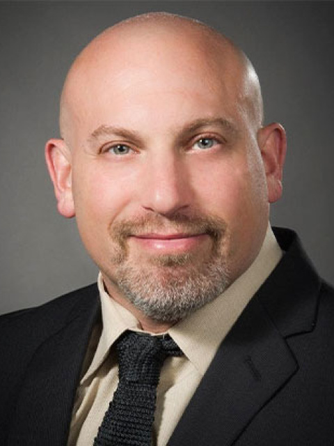
Dr. Seth Koenig has been interested in bringing point-of-care ultrasound to critical care for more than a decade. His efforts and those of his associates and colleagues have helped revolutionize the way critically ill patients are managed in the U.S. and around the globe.
Dr. Koenig’s research interests have led to numerous studies and publications involving point-of-care ultrasonography, particularly in critical care.


This educational webinar is intended for healthcare professionals and not for patients or consumers. The material is provided for general educational purposes, as a reference and a supplement to professional experience, education and training, and should not be considered the exclusive source for this type of information. The presenter in this webinar is a paid consultant of FUJIFILM SonoSite, Inc., and is sharing the presenter's own views and opinions that do not necessarily represent the views and opinions of FUJIFILM SonoSite, Inc. This educational webinar is not intended to recommend any device for a particular indication or to provide indications for use for any device. At all times, it is the professional responsibility of the practitioner to exercise independent clinical judgment in each particular situation. Fujifilm assumes no responsibility or liability for any misuse of the information imparted in this webinar. This educational webinar does not supplement, replace, or supersede device labeling, including instructions for use, which accompanies any FUJIFILM Sonosite product.