


Transcript
- My name is Chris Pennell.
Welcome to the Fujifilm Sono site
behind the Scan webinar series.
This webinar is entitled, developing
and Maintaining Skills for TEU in Acute Care.
Before we begin, please be advised all attendees are muted
and you can type your questions into the q
and a box in the toolbar located at the bottom
or the site of your screen at any time,
and we'll conduct the q
and a session at the end of the program once Dr.
Tarran is finished with his presentation.
This webinar will be recorded
and will be up on sono site.com afterwards.
It's my pleasure, pleasure now to introduce our moderator,
Paul Bosky from our clinical market development team.
So Paul, I'll give it to you and you can take it away.
- Thank you very much, Chris. I appreciate it.
On behalf of the entire Fujifilm CY team,
it's my esteemed pleasure to welcome you all here today
to the first in a series
of webinars on trans esophageal ultrasound.
Today's topic, developing
and maintaining skills for TEU in acute care.
And today's guest speaker is Dr. Felipe Tarran. Dr.
Tarran is an assistant professor of the Department
of Emergency Medicine at Weill Cornell Medicine in New York,
univer, New York City,
and he's a leading expert in emergency
and critical care ultrasound.
His resource focuses on the study of strategies
to improve outcomes in cardiac arrest
and resuscitation, specifically incorporating the use
of resuscitative transesophageal echocardiography
or TEE.
He is the founder, is of the resuscitative TEE project
and is the principal investigator
of the multicenter TEE collaborative industry.
As the course director for the Resuscitative
TEE workshop, Dr.
Tarran leads a group of multidisciplinary experts
and has helped train over 1300 physicians in the
United States, Canada, and Australia.
Those courses have included courses at the Society
of Academic Emergency Medicine,
the Emergency Medicine Residents Association,
and the World Interactive Network focused on critical care,
critical ultrasound, otherwise known as Wind Focus.
Dr. Tran, it is my pleasure to welcome you here today.
We look forward to a informative, robust,
and interactive presentation,
so I'm gonna hand it off to you.
Thank you very much.
- Thank you very much Paul,
and thank you to the entire Sonos Set team
for having me for this opportunity.
The format, if, if it wasn't explained, we're going
to do a discussion.
We're gonna have a, a brief presentation on this topic,
hopefully no more than 25 minutes.
And the idea for this live webinar is to have most
of the time we have available to actually have
the opportunity to discuss questions,
live questions from the audience, as well as some questions
that were submitted ahead of time.
So the, the, the idea will be to
hopefully use those questions to further clarify some
of the important nuances and,
and topics that aspects of this, of this topic
that are relevant for your practice.
In this presentation specifically, I'm going to share
with you my personal perspective, obviously on this topic,
and it's the perspective that I acquired over years
of teaching fellows, teaching residents,
teaching faculty in while implementing this modality TEA
in several emergency departments.
It is also when my team
and myself have learned in the process
of training over 1200 clinicians, both emergency physicians,
intensivist, and an ologist
through the resuscitation tea workshop.
This international course created specifically
to teach physicians the fundamentals of this modality
as a new modality in point of care.
Ultrasound T is increasingly being utilized in emergency
departments in ICUs, unlike surface ultrasound
where we can easily practice in either standardized patients
or volunteers or simply amongst ourselves when learning.
An important issue with trans esophageal echo is that
because of its invasive
or somewhat invasive nature, just like with surgery,
we ideally need to have access to simulation training
where we can develop those necessary skills
to practice this modality, not only safely,
but also efficiently in the clinical setting.
So when we talk about training in transesophageal echo
or trans esophageal ultrasound, we need
to consider the scope of practice.
Define very clearly what we are talking about here.
We're talking about the performance of focus
or resuscitative or rescue
or TU in the emergency or critical care settings.
That is T
or TU performed at the point of care by clinicians
or the clinical team caring for a given patient.
And the exam is driven by a specific clinical question.
So this is the, the most important difference from
comprehensive or consultative T.
So there's going to be a specific clinical question at hand
or a specific application
for which we're using this modality.
So this could be evaluating a patient during cardiac arrest
as we do, doing what we call T guided CPR
or T informed CPR to evaluate for reversible etiologies
to optimize the quality of chest compressions
or characterizing the etiology of shock in 2022.
We'd like to say that there shouldn't be
th there shouldn't be patients that we treat
for a long period of time as undifferentiated shock.
We have the technology, we have the modalities to establish
fairly accurately what is driving patient shock,
what are the underlying mechanisms, and T
or t, U is one of those modalities
that can help us accomplish that objective.
Same with guiding endovascular procedures, A
of endovascular procedures, VA ecmo, VV ecmo, impellas
balloon pumps, tps.
A number of procedures that we do in the setting
of critical patients can be optimized, can be improved
with this, with this mortality.
And while this means that we are in most cases
using only a few views of TEU in order
to obtain the information that we're looking for to answer
that specific question
or that that set of questions, the number
of views is really not what defines the practice of TEU
or resuscitative T, it is the scope, actually the question
that we're asking the emergent nature of the study
and the fact that it's the information that we're obtaining
by definition should impact the decision making at the point
of care in in real time.
This is an important point
that I feel often gets missed when we talk about TU.
And it's also an important point as we define competency
and talk about TTU and emergency settings.
Yes, in order to perform focus TU and emergency department
or in the ICU setting, you need to know only somewhere
between four, maybe six, eight
or up to 12 views
as we teach in our course depending on the application
that you're using specifically.
But really the truth from a competency standpoint,
this is a message that I want to convey
during this presentation and during today's discussion.
From a skills perspective, you just like swimming
where you either swim or you don't swim.
There's just no in between.
With T you, you either know T or
and understand it as a modality or you don't.
Therefore, in this presentation, what I'd like to
to give you is a framework to think about T training.
We will review what are the key motor
and cognitive skills required for for this modality,
the critical role of simulation training for T, the pros
and cons of most
of the common experiential learning environments.
For instance, the operating room
with our colleagues in cardiac anesthesiology,
the echocardiography lab with our cardiology colleagues
or the emergency department.
So we can actually practice T
and teach T with those cases
that we perform in in our department as well as in the ICU.
We're also going to touch on the evaluation
and the importance of assessing competency in skill
maintenance in in focus T,
and hopefully we'll have a discussion involving
those aspects as well.
Let's start by discussing the skills required
to competently perform te.
How do we get to procedural competency
to procedural proficiency?
Competency in resuscitative tea requires the development
of two different types of skills.
Of skills, rather mortal skills
that is manipulating the probe obviously,
and the different controls.
We have to understand the knobs, understand how they work.
Just like when we learn how to play a guitar, we want to get
to the point where we can play the guitar not having
to look at our left hand if you're right-handed
and look at the chords,
but rather know how that feels
and know exactly where we need to press and when.
So similarly, the within the motor skills in T, we need
to really become familiar
and become one to some extent with
that control in its mechanical and and digital controls.
And the first component of these motor skills, just kind
of dissecting this broadly, is going
to be probe insertion, right?
We need to insert this probe in order to generate images.
And this is often actually a component of training in TE
and tu that is, I think, under appreciated
when you develop the ability to quickly
and safely insert the probe in the setting
of an intubated patient.
In many cases, like the case of cardiac arrest specifically,
we need to perform this procedure under pressure.
We're doing it while the entire team is looking at us
waiting for that information,
hopefully not only within a few seconds,
but also we want to do it without interfering
with other resuscitative interventions.
For instance, airway management, the chest impressions.
So in resuscitative t in general,
patients are always intubated as part of, again, the scope
of practice of this metallic, and
therefore this portion
of the procedure follows advanced airway management.
In this picture here from the T workshop,
you see a physician practice in probe insertion technique in
a prototype training that we, our team developed
for this specific purpose.
And as part of the skill,
understanding the anatomy is actually a key aspect
of development.
The probe insertion proficiency, understanding
of the anatomy that is relevant
to a te probe insertion is really key.
And it is key because the most common anatomical sites
causing obstruction of the TE probe during the insertion
and in intubating patients are the adenal cartilages
and at the end of the hypopharynx, the filosa.
So when we teach this
and the, the, the primary goal here from a competency
standpoint is going to do, is going
to be maintain midline as we descend down
to the base of the tongue.
And then at that point we need to do a maneuver to lift,
to displace the esophagus anteriorly.
So in order to access the esophagus and,
and avoid pharyngeal injuries,
which I think are under appreciated, it is really critical
to maintain midline while advancing the probe
down in the hypopharynx.
This can be performed either digitally guiding the probe
just under with tactile confirmation with your fingers.
This is what I often do
or under direct visualization with video endoscopy.
At this point, we actually have a number
of studies including a recent randomized control trial out
of India that demonstrated
that it is actually more efficient
and safer in terms of the rates of,
of pharyngeal injuries to guide this procedure
with laryngoscopy and specifically video laryngoscopy.
But you have to know the anatomy, you have to know that,
for instance, the esophagus is most
of the times in humans slightly off to the left.
And this, this is the rationale
to secure the endotracheal tube
as you see in this picture on the right side
and go with the end with the probe on the left side.
So this is going to be the first of those motor skills
to aid, as I said, the passage of the probe, the,
the probe's tip past that upper esophageal sphincter, the,
the mandible, the jaw needs to be pulled forward gently in,
in the patient's neck, slightly forward flex.
So, so this is going to be different than
when we're attempting to intubate in
that we are extending the neck.
In this case, just like with an OG
or an NG tube, we're slightly, we're providing slight
forward flexion or flexion of the neck.
So while advancing the probe, it is really important
to be cognizant of the endotracheal tube.
One of the complications that we are tracking
that we're looking for in the resuscitated T collaborative
registry, the multicenter registry,
collecting data on the clinical impact
and outcomes of, of this modality.
One of those complications
that we're tracking actually is precisely
dislodgment of the endotracheal tube.
This is something that has been brought up repeatedly
over the years, however, until up to this point in, in,
as we presented at asip recently,
more than 500 cases that have been done at that point of
that interim analysis that there had been
no dislodgements of the tube.
But it's important to understand these anatomical aspects,
both again, to prevent injuries as well
as complications such as dislodging, the tube as well
as doing it efficiently.
And the second component of these motor skills, again,
kind trying to dissect this so that we can work on each one
of these components.
And this is exactly what we do during training
of our trainees as well, as well as during the workshop,
are the skills that are required for image acquisition
and to develop the view.
So just like with any procedure
that involved hand-eye coordination, we need
to develop the skills
to control the different movements of the probe.
In this case, there are mechanical movements
that include the rotation of the probe left
and right, advancing the probe, drawing the probe,
and then flexion and retro flexion, the flexion forward and,
and inflection backward at the tip of the probe
is, is key.
And we need to also become familiar
with the digital movements.
This is something that creates often
confusion amongst learners.
There are mechanical
and digital movements with this modality.
The digital movements are those that are
provided by the OmniPlan.
The OmniPlan is essentially the ability to rotate the beam
of the TE probe that really defines tea.
It is really what makes TE the powerful tool that it is.
So in integrating this, different movements
takes deliberate practice.
It is important to first understand how each
of these movements work individually
and then learn how to integrate them.
This is again, just like what we do with other procedures
that require similar coordination, similar type of skills,
and it's also the what we should be doing,
what we learned transthoracic.
And this is the part where it really makes sense to utilize
high fidelity simulation to learn these cont these concepts,
these controls to develop the tactile feedback
of these different movements.
Again, to be able to do this without having
to look at your left hand
or right hand, depending on if you're lefty
or right-handed, particularly those mechanical movements,
for instance, like flexion of the probe, which
as you might have thought already flexion of that tip
of the probe in the esophagus is
what could potentially cause injuries.
And therefore we need to be very cognizant of, of, of
where we are, how much we're flexing,
and that is best achieved in, in the setting
of high fidelity simulation
before we start practicing a real patients.
The other type of skills required
to perform TA proficiently are going to be cognitive skills.
The skills that we need to develop images to,
to interpret the images, rather to identify both normal
and abnormal anatomy as well as the pathology
that is gonna be relevant to these studies.
This is where emergency physicians
or intensive care physicians, anybody really in acute care
already have experience in focus.
Transthoracic ultrasound
for the most part is really a standard in the
emergency department setting.
And therefore we as, as a group of physicians, as a group
of practitioners, have a significant advantage when
learning tea.
Many people don't really know this,
but in anes sociology until just a few years ago
before point of care became,
before ultrasound became a core competency
of an anology training
of residency training fellows in cardiac cardiothoracic
sociology would actually get to the OR in July beginning
of the training and be first exposed to TE having a minimal
or simply no previous knowledge of ultrasound,
and they would literally learn te from scratch.
And this is, it was very shocking, sort of interesting to me
as a learner many years ago when I first spent time
with my mentor in the, in the operating room to see that,
that that was the pathway of, of an anesthesiologist
to learn in that case advanced applications
of perioperative te.
So emergency physicians and ICU physicians,
and again, anybody in the acute care setting
and an anesthesiologist practicing also non,
non in a non-operative setting have really already a leg up
when it comes to learning t for this reason,
they already have knowledge of the anatomy
of the structure seen in some of these T images
that are essentially mirror images
of those in transthoracic.
And so the challenge,
and this is what's often underestimated in my opinion,
is really to understand the cardiac structures
and the anatomy from this novel perspective,
from the retro cardiac perspective.
And if anybody in the audience has attended our workshop,
we, we have really defined this as one
of the defining principles of our own approach
to transesophageal echo
and the curriculum that our team has developed
and refined over many years of the workshop.
We spend really dedicated time during course
and during this training, teaching trainees this novel
perspective of the heart.
So we ask people to essentially rewire their brain,
to visualize the anatomy from this different perspective.
What is the probe seen from this retro cardiac position?
If you develop the ability to understand that
where the imaging planes are cutting the heart in the great
vessels, you can interpret t images in a way
that it doesn't really rely on memorizing image patterns
as most people, I think unfortunately intuitively do when
learning tea, they just go to a textbook or an atlas
and they learn 35 degrees, 35 centimeters of,
of depth in the esophagus,
and then I'm going to rotate OmniPlan to X degrees.
Unfortunately, they try to memorize
what they're really supposed to see in HVU.
And what we've learned through our experience is
that it's much more beneficial
and again, leads to a more durable, developed
leads to more durable skills and competency when physicians
and practitioners in general actually understand the anatomy
from this novel, again, this retro cardiac perspective.
And while developing the skills might seem daunting,
might be maybe overwhelming to you
and I, by no means I, I want
to make t make t sound more complicated than it is.
We want to make it as simple as it can be to,
to train physicians on this modality.
I've actually got really good news for you
as an emergency physician
or again, intensive care physicians,
anybody working in in in acute care settings,
we've been really training for TE for years.
Even if you didn't know that you were training for te
because TE really builds on existing motor
and cognitive skills
that are really central to our practice.
That hand eye coordination, those kinesthetic abilities,
that is the sensory perception of movement.
Those are really at the core of procedural training
of emergency physicians and critical care physicians.
So think about ultrasound guidance of procedures,
aary management maneuvers, vl, NPL as shown in this case
by one of our residents.
Our proficiency in those procedures provides us
with a really strong procedural foundation to learn
and perform t.
So we're really in a good, in, in a good position,
again, as learners to to, to add this modality
to our, our momentum.
So having to find what those specific skills are
that we need to acquire, that we need
to work on when training in, in t
and TU, let's now review the specific
and critical role of high fidelity simulation.
As I told you in the beginning,
I think critical high fidelity simulation
as seen in this picture, one of the hard work simulators
by intelligent ultrasound com, that simulators
that we use in our doing our trainings.
This is really I necessary tool, I believe not only
for access for development of competency,
but also for maintenance of competency.
In some settings, a comprehensive review
of the literature
or of the evidence in this area really is
beyond the scope of the sessions.
However, I want to briefly describe to you some
of the key theory.
These may be some examples that I think frame some
of the theories supporting the use of this, of this modality
of, of simulation high fidelity simulation for,
for training in this, in this modality specifically,
this is one of the studies for instance, by block
they evaluated 44 ICU trainees fellows
and then randomized them to either lecture-based curriculum
or lecture plus
for our structured curriculum and simulation.
And as you would expect, again, this is just
to give you an example of what I think is obvious,
as you would expect,
simulation-based training improves the ability
of novice operators
to perform a focused critical care exam in this case
as the scope that they had defined for that study compared
to a lecture only based curriculum.
And this is again, what you would expect.
Chen and Tom Angelic, one of our faculty and
and members of the S project did a similar study in Canada a
few years ago in this case with emergency physicians.
Again, very similar
and I encourage anybody who's interested in,
in the literature, we have several
of these studies on their s tea projects website under the
section of curated literature in this case,
they reported the success rates for each specific view
and each structure that that was being evaluated.
So recommend that you check that out.
Again, a as as predicted, they demonstrated that simulation
works that we can teach physicians
with a structured curriculum
and that that leads to the development of those motor
and cognitive skills that I just described.
Other more sophisticated studies like,
and this one performed by a
or on a group of cardiology trainees, cardiology fellows,
if I remember correctly, evaluates motion analysis in
how training with simulators actually impacts
proficiency in t.
This is something that I've,
I've always found that are interesting.
And if you think about it, it makes a lot of sense with,
with simulators, high fidelity simulators,
just like hard work simulator, they're essentially
very powerful computers.
And so we can actually not only assess the trainees
externally in terms of visualize observing them
what they're doing, but we can actually track the data.
We can, we can evaluate from the computer side
of things how efficient
or not efficient they are when developing those views.
So this is what they did in this study.
They used a multimodal training curriculum
that include high fidelity simulation.
And as you see in this graphs here,
this is again a kinematic analysis of of
of transesophageal echo training in cardiology fellows.
If you see the figure there, B versus C,
those are essentially the tracings, the three tracings
of expert compared to novices in terms
of their, their, their, their curve, their learning curve.
So the point here is that as again, you,
you one would expect you begin as
shown in, in, in picture
or in graph number A,
it is very inefficient.
You are kind of erratic, you do a lot
of inefficient movements.
You're wasting seconds
or fractions of seconds trying
to get to where you're growing.
And as we develop more competency on the simulators,
we become more efficient
and becoming more efficient not only means getting,
developing the view, but actually means getting the view in
a more efficient way faster,
which if you think about not only represents potentially
just getting the data that you need faster,
but also in the case of this invasive modality,
it represents less risk for injuries.
If you have a cleaner,
just like think about video laryngoscopy
or laryngoscopy in general, if you have a cleaner
descent down to the
or pharynx to, to perform your epiglotoscopy
as a a a compared
to somebody entering not really understanding
what they're seeing janking the, the laryngoscope back
and forth or moving it laterally, each of those sort
of erratic or non, non
not needed movements is going to represent again,
potential for injury.
So what the, this study
and other similar studies have demonstrated
with the sch nomatic analysis is
that there is actually not only an improvement in the
ability to develop the views,
but also an improvement in the,
in the proficiency of developing those views as far
as getting there in a more ergonomic and efficient way.
And while you might think at this point, well, I need one
of those very expensive high fidelity simulators and,
and that's really all all we need for training,
I actually want to supplement to that perspective,
that point with another of the most important really
lessons that we've had from our experience training hundreds
of physicians in this modality.
And that is a in, in a, in the ideal training
for physicians.
And I, I would argue that this applies not only
to any modality of ultrasound,
but I think to any procedure in general,
multimodal learning is really I think the best approach this
data supporting this, what it means is in the, in the case
of Transvaal Echo, for instance,
we're actually using analog methods,
in this case 3D printed models
to actually ha help people understand this difficult
anatomical concepts that I was alluding to in the beginning.
Understanding what is the probe scene from the, from
behind the heart, what are the different changes in
situation and position of the heart within the chest
and how we go about developing the view.
So it might seem counterintuitive.
Why are you using this hard models?
If you can see this beautiful 3D models in the computer,
in the reality, in both our experience as well as, as again,
studies have shown that there is actually great value
that our brain really benefits from having input in the form
of different modalities.
So we're gonna have people train, learn the theory,
consume materials online, their own pace,
digest some images, read about cases,
then we're gonna compliment
or supplement that with high fidelity simulation training
to teach them really the, the, the fundamentals of the,
of those motor skills.
And then in, in addition to that, we're going to supplement
that cognitive part that is
provided by the high fidelity simulation
with analog techniques such as this work, what we use
for instance, vectors to understand the plane,
to really help our brain sort of break this complex
skills down into chunks that we can work on
among other things in technologies that we've incorporated.
The use of augmented reality in this, in this case
where you see here is a tablet from Hardwork called Hardwork
AR is essentially similar software than the one that is
loaded in those high fidelity simulators.
In this case, it is something we can actually hold in our
hands to e tablet allows us to cut
through the hard in the different planes that we use
to develop those T images.
And again, this is another example of
that multimodal approach to training,
supplementing again the training in the simulator with
these different modalities.
I wanna wrap this up by giving you
a few points on the pros
and cons of the most common
experiential learning environments.
What we mean by that is essentially emergency department,
for instance, would be an option as I mentioned,
just learning from tes that we perform in the ed,
the operating room would be kind of the natural,
probably the most common is experiential learning
environment for TEU in the setting of,
of perioperative te specifically.
So I wanna give you my perspective on
what each of these environments can give us
and why you should consider, for instance,
getting access to the operative.
In my opinion, in addition to simulation,
the operating room is really the ideal training environment
for us as emergency physicians or intensivists to train.
This is why we advocate for alliances,
for partnerships with our cardiac
and theology colleagues to get access
to the operating room setting and gain experience with t.
Some of the advantages
of training t in the operating room include
that it is a controlled environment.
The patient is in perfect positioning.
We have monitoring, we had the direct supervision in a
controlled environment by an experienced faculty,
in this case a cardiac anesthesiologist as anesthetist,
you can practice probe insertion,
which is something we can't quite do in the simulators
as I showed you in that image earlier.
We use a trainer to practice that,
but obviously there is, you can't compare the experience
of actually inserting the probe on a real patient.
So after developing skills, for instance on the trainer,
you, you should have some experience again with guidance,
with somebody looking over your shoulder to,
to learn how to insert the probe.
These are again, key mortal skills that need to be
developed in that, in this case,
in the operating room can be developed very efficiently.
So it is a control environment.
We have the opportunity to practice probe insertion.
You can spend hours really scanning the same patient.
This is something that I tell my resident,
my fellows cardiothoracic surgeries
last 4, 6, 8, 10 hours
and the prob stay stays in for the entire case.
So before they go and bypass, after they go
and bypass during bypass,
you can use all those opportunities to scan
to learn the anatomy for a sociology fellows, in fact,
I learned recently that for purpose of counting their scans,
the scan that they perform pre bypass
and the scan they performed on the same patient post bypass.
When they come out of bypass, a critical part of the,
of the anesthesia is actually considered,
they are considered as separate scans
of separate studies towards their 300
or one 50 depending on the, the scope of basic
or advanced perioperative.
T The other aspect that is really unique
and advantage of training in the operating room is
that you can see pathology.
We have the opportunity to see pathology
that we might either not see ever in the emergency
department or that we might see infrequently depending on,
again, where you work.
Just as an example, when I was a trainee,
I would have my mentor to actually
text me when she was about to do a study
or she knew that he was going to do a study that day
for a case where there would be pathology to see so
that I could gain the experience of, of evaluating, say,
aortic dissections that were being repaired
or valve pathology, massive PEs going for thrombectomy
VA related complications, ecmo, cannulation,
impella placements, so on and so forth.
So these are kind of pathology
and in applications of TE
that you might just see in frequently in the emergency
department setting or the ICU setting and the eyes
and the, the elective or emergent setting of the,
or really represents a unique environment to, to do that.
I cannot recommend enough spending time in the operat room
for that reason, unlike cardiac anesthesia or cardiology.
However, for us, as I
briefly mentioned in the emergency department setting
and also in the ICU, again, depending obviously
where you're practicing, there might be some variability.
The frequency of studies is going to be relatively low.
Therefore, access to simulation, I believe as I mentioned,
is critical not only to
for skill acquisition,
but also for maintenance of competency.
There are currently three comparable simulation systems
market in the us maybe four actually at this point.
And all through all use virtual reality
with digital reconstruction to generate t images.
So all offered packages with clinical cases
with online version of the software.
And in sake of time, obviously we don't plan to cover it,
I don't plan to cover here each one of these systems,
but you can get information on these different systems also
again, online through our website.
But in order to summarize again
how you should think about if you're trying to cons,
if you're considering simulation
or you're, you're asking yourself why is, is this important?
What, what, what does it bring to the table, if you will,
it allows deliberate practice.
That is something we can't quite do when we're
scanning a real patient.
We can't just like stop
and think about one aspect at least is not as easy
as it is with, with a simulation.
So we can break down these complex skills into
chunks that we work specifically
for an allocated period of time.
It is really the most efficient way
to build a foundation in transesophageal echo.
It is a unique tool both to learn
and to teach anatomy in that spatial orientation
that I told you that is critical to understand
how these images are generated.
It can be adapted to learner needs.
We can make it as simple as needed
for a learner depending on where they start.
This is exactly what we do during our workshop.
Again, we start, for instance, working
with simulation using deliberate practice
to only understand the,
the anatomy using the 3D images without the 2D images on
purpose so that we can build that mental map first
and then move on to the 2D image interpretation.
Fidelity simulators have haptics
that really, that are incredibly good, that,
that make those model skills that we acquired
really highly transferable to patient scanning.
This has been really surprising to me
and the, the way the technology has evolved over the years.
In other words, after spending time again with deliberate,
with, with structured training on simulator,
not just doing whatever you want, not just kind of playing
around, but rather having a structured approach on this
powerful systems,
you can actually then transfer those skills you'll be ready
to, to rep, replicate
those studies in real patients.
And lastly, as I said, I think as an argument, it really has
an, a unique role not only developing those skills,
but also maintaining proficiency
and competencies specifically in emergency medicine.
If you think about the data we have from our registry at
this point and from previous surveys,
most centers are doing anywhere between five
or just a handful of studies per month
to maybe 20, 25, 30 in some
of the centers that are doing the most.
So this is really to some, if you divide that, the,
if you then divide that up in whatever number
of faculty they have in that center doing tes, you are left
with just maybe one or two
or less than one study per month by physician.
In some cases you can go for months not having done a study
after developing your, your sort of foundational training.
And I don't wanna make it sound like this is a rare scenario
for us in emergency medicine.
We are all expected to perform pure mortem
or resuscitative c-sections.
We're all exploited
to perform cricothyroid autotomies in the setting
of error management.
We're expected to do cla shells
or thoracotomies and none of these procedures are frequent.
And so some of these problems are actually common to us
and we, we have dealt with them.
But similarly, TI think,
and this is I think a key role for simulation, again,
it is an opportunity for us to get a refresher to get
those mortar and cognitive skills kind of back
and brushed up every, every, every so often.
So I think again, not only for development of competency,
but also for, for maintenance of those of those skills.
Just to be practical, for instance, in our institution,
we're including the use of simulation
as in proctor studies on a simulator as a,
a way to get to, to maintain competency
and to, to maintain privileges once a year.
So lastly,
I wanna just touch brief very briefly on the aspect of,
of competency assessment in TU.
How many studies really do you need?
And this is going to depend obviously in your environment,
whether it's in emergency department or ICU,
but I want to leave you with some numbers.
It is well known at this point from critical care literature
that it is somewhere around 30 studies, 30 to 36 studies
in the case of advanced critical care echo competency.
So this is actually a different scope than the scope
of practice of TU and emergency medicine,
at least the the basic one,
this actually involves hemodynamics, involves,
involves identifying severe bowel pathology.
It is what in, in currently in the United States is now
formally sort of described and,
and contained within the advanced critical care
board certification.
So for that advanced critical care, echo board,
critical care, echo scope for physicians
that had prior competency in transthoracic,
it took roughly 31 to 36 examinations to get them to
that point of, of proficiency developing
and interpreting views.
A number of studies, some of the citations here, again, most
of these citations and others are in,
you can find them in the ity
projects website under the section
of literature in the emergency department.
We have a couple of studies at this point in the, again,
emergency department setting specifically,
and the number is somewhere between 10 and 25 examinations.
So using a focus proc focus, I'm sorry,
a structured curriculum that that involves the use
of high fidelity simulation.
We know that it is feasible to teach physicians the fun,
the fundamentals of, of TTU
and get them to a point
where they can develop those core views and interpret them.
So again, a number of these studies have been demo,
have been published in the last few years.
It is important to recognize this caveat here
or this requirement again
with prior competency in transthoracic.
So for somebody that already has that level
of competency on top of that, if we have
that structured curriculum, somewhere between 10
and 25 exams will get us where we need to be.
And this is the, the number that very, again, generally
ASEP recommended in the guidelines published in 2017,
where they actually recommended 10 proctor exams
as the number sort of needed to develop competency.
And again, this will vary in across institutions,
especially when it comes to privileges, which is
by nature a institution dependent process.
With that, I want to thank you for your attention.
This is my email Cornell,
otherwise the website has a lot of resources
and I would love and
and look forward to taking any questions from, from you.
- Dr. Duran, thank you so much
for a very informative presentation.
I, I will remind people attending
that we have opened up the q and a session.
If you just choose a q and A bar across the middle,
you can enter it in and we will monitor
those and ask them to Dr.
Turan. Doctor, I had a couple of emails that were sent
to me separately with questions.
If, if it's okay by you, you quote,
you spoke quite eloquently about the access to simulation.
If you don't, if if someone doesn't have a simulator,
if they don't have a sim lab, what is the best way
to go about getting access to simulation training?
- For sure. That's a great question
and a question that comes up all the time when we interact
with physicians that are interested
in this, in this modality.
It just had this question probably asked me a number
of times at asep as, as you saw in this presentation,
I personally and my team believes that simulation is key.
That is the best way to get us
Trinity safely and efficiently.
But it's a reality that these systems are expensive,
that they're over a hundred thousand dollars and most cases.
And, and it's safe to say
that not every emergency department
or not every A CU will have access to one sort
of dedicated system for them.
This is exactly why our team, the Resus
project developed a course
years ago in 20 17, 16, 17,
to provide physicians that
otherwise wouldn't have access to simulation with
that opportunity to get simulation training
under a structured curriculum.
So obviously, and the, I i I need
to acknowledge the conflict here as a course director
of this workshop, the workshop that we run, other courses
that you might find out, there are one option that you have
to get ex exposed to get simulation training.
Beyond that, there are many simulators that are owned
by simulation centers,
especially simulation centers within hospitals
that the central kind of entities
that often are accumulating dust,
sitting somewhere not really being used.
And this is something that unfortunately we have
learned also over the years.
You need something else than just the system.
You need a curriculum, you need somebody
to actually teach you on that system.
And so in the, in the case of those simulation centers,
in many cases, I think emergency departments
or IC can get access to those
and bring experts from their team from elsewhere
or set up something with their cardiology department,
with cardiac anesthesia
and have the opportunity
to learn on one of those simulators.
Again, simulator simulation centers often have
them, and you might not even know
that your simulation has center has one as a third option.
I think regional training, the idea to have some simulators
that can be shared within hospitals
or by different hospitals within a region is something that,
I don't think it exists today,
but I think it should be a goal for us as a specialty.
For instance, this is example
of endoscopy training centers, for instance.
This is how endoscopy became
a widely available intervention within gi Many years ago
simulators were, were not widespread,
were difficult to get access to, and
therefore there were centers that became sort
of training centers for this modality.
And I think something like that regionally, nationally,
would make sense for this modality in emergency
medicine and critical care.
But ultimately, if you don't have an option to access
to a simulator, but you have access to an operating room,
you have colleagues, you have a cardiac thoracic, anes
department division
and program, I think getting access
to the OR would be the next, the next best option if, again,
if you can't start with simulation, I personally believe
that it's not ideal to start from scratch on humans
that we should get, you know, the first part
of our learning curve in, in a simulator.
But if you don't have a, it is the reality
that in many centers,
the first exposure takes place again in the setting
of the operating room.
- Sorry, we have a couple of questions from our attendees.
Jonathan wants to know, a, as you know, ultrasounds,
US scopes are more common than echocardio scopes.
Do you think that there is any benefit to supplement tte
and if it translates to resuscitate of TEU
- Ultrasound scopes are more common than echocardio scopes?
I don't think I understand this question.
Do you understand the question, Paul?
- I, yeah, I had a little bit of a tough time with it.
Jo, Jonathan, if you'd like to clarify
- That.
Sorry, Jonathan, I I'm sure it's me.
I am, I'm not following you.
Do you think that there are, is any benefit
to supplement TTE
and if, if it translates to resuscitative TU?
Yeah, I'm not following it. I know this is
Jonathan, it's a CNA.
I would love to understand this question.
If, if you can maybe provide us with a,
a little bit more information there in the,
in the chat, would love to answer it could do that.
- We would, yeah, we would appreciate that.
The next question in, in full disclosure here,
Fujifilm Sono site is the exclusive ultrasound sponsor
for the resuscitated TEE workshop
and Dr. Tran, when we have your 2023 schedule
of courses out for the coming year.
- Yeah, that's a great question.
So this is as a surprise actually.
The, the, the calendar will be released tonight,
and if you go to the website tonight, you should be able
to get to the, to get the calendar, we're gonna start
in April.
In Miami will be April 8th will be the first course,
but there'll be a number of other courses
just like previous year.
So if anybody's interested, actually tonight
after around eight, 8:00 PM e eastern time,
you should be able to see the calendar and,
and everybody who's signed up
to on the website will receive notification.
- Okay, very good. Thank you.
Another question that was emailed earlier,
what are the most common mistakes you have observed
among physicians trying to learn TEE?
Any tips you can offer them?
- Yeah, so there, there are a number obviously,
and I think I I touch on a, a few of these,
probably the most important one that I've seen in that,
that I think remains unfortunately a recurrent problem.
This is why we, we are so deliberate about this
during our training, myself and,
and my institution with my trainees and,
and again, our team at the, at the workshop is the,
the this in intuitive approach to learning te in a way
that we just memorize images, that we memorize kind
of the recipe, the sort of cookbook recipe of how
to get to the image.
And unfortunately the reason why that happens is
because simulators specifically are
developing the images on a simulator is fairly easy.
If you follow that recipe, if you follow the, you know,
depth within the esophagus, then the OmniPlan rotation,
if there's need to rotate at all,
we'll get you to the right view.
Unfortunately, what happens with real humans is that
unlike the simulators that have a normal anatomy
or that represent a normal heart, is
that humans have a lot of variability in the anatomy.
And if you think about it, the patients that we scan,
the patients in whom we need to obtain data with a TU
in the critical care emergency setting are patients that
by definition in most cases will have chronic pathologies
that that led them to the acute event
that we are treating them for.
And so the fact that patients
that we scan will have dilated cardiomyopathy,
right ventricle hypertrophy that will have holcomb,
that will have pathology will required from us
to understand that anatomy
and to adjust this, this sort
of cookbook recipe directions from, from the textbooks,
from, from whatever we've learned and, and, and,
and modify them to actually develop the views and, and, and,
and in our individual patients.
So that is really one of the most common mistakes
that I see is, is unfortunately intuitively the way we learn
TTE is that we memorize images as we memorize kind
of the pattern that we're looking for.
And you can, you can get away with that in transthoracic
because there's only limited places
where you can put the probe.
Unfortunately with transesophageal, there are multiple,
almost unlimited possibilities of, of positioning that probe
and then rotating that, that beam and
therefore that option just doesn't work.
We can't really rely on that, on that memorization of,
of image patterns.
We actually need to understand what we're seeing.
So I'd say that's probably the most important
mistake that I see in terms of,
of training and approach to it.
- Okay. Excellent. Well, Dr.
Teran, it looks like our questions
and answers are pretty well used up
and our attendees are slowly drifting off.
I want to thank you again for taking part in today's and,
and, and putting on today's webinar.
It was most, most educational
and I encourage folks who are on, if they would like to
or signed on late to it, if they would like to access this
or maybe pass this on to any of their colleagues
to please go to sona site.com and search under webinars
and you'll be able to find out information there again
and a recording of this webinar there.
Again, Dr. Tren, thank you very much for your time
and I encourage also the folks who are here to look
for the next in our series
of trans esophageal ultrasound webinars
that we are working on for the end of January, 2023.
Take care everybody, we'll be back. Thank you.
- Thank you.
Transesophageal Ultrasound (TEU) has evolved over the last decade and has become a valuable tool at the point-of-care. Education and training are essential when learning the skills necessary for the use of TEU at the point-of-care. This webinar will discuss developing and maintaining skills for TEU in the Acute Care environment.
What You'll Learn
- Key motor and cognitive skills required to perform TEE
- The role of simulation training for TEE
- The Pros and Cons of the most common experiential learning environments for TEE; Operating Room, Echocardiography Lab, Emergency Department, & ICU
- The evaluation of competency and skill maintenance in focused TEE
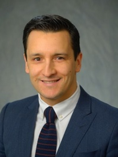
Dr. Teran is an emergency physician and investigator at Weill Cornell Medicine in New York City. He completed a fellowship in Emergency Ultrasonography at Mount Sinai Hospital and a Master of Science in Clinical Epidemiology at University of Pennsylvania. As a leading expert in emergency and critical care ultrasound, Dr. Teran’s research focuses on the study of strategies to improve outcomes in cardiac arrest resuscitation, specifically incorporating the use of resuscitative transesophageal echocardiography (TEE).
He is the founder of the Resuscitative TEE Project and principal investigator of the multicenter TEE Collaborative Registry, aimed to accelerate the development of outcome-oriented research and knowledge translation on the use of TEE in emergency and critical care settings.


This educational webinar is intended for healthcare professionals and not for patients or consumers. The material is provided for general educational purposes, as a reference and a supplement to professional experience, education and training, and should not be considered the exclusive source for this type of information. The presenter in this webinar is a paid consultant of FUJIFILM SonoSite, Inc., and is sharing the presenter's own views and opinions that do not necessarily represent the views and opinions of FUJIFILM SonoSite, Inc. This educational webinar is not intended to recommend any device for a particular indication or to provide indications for use for any device. At all times, it is the professional responsibility of the practitioner to exercise independent clinical judgment in each particular situation. Fujifilm assumes no responsibility or liability for any misuse of the information imparted in this webinar. This educational webinar does not supplement, replace, or supersede device labeling, including instructions for use, which accompanies any FUJIFILM Sonosite product.