

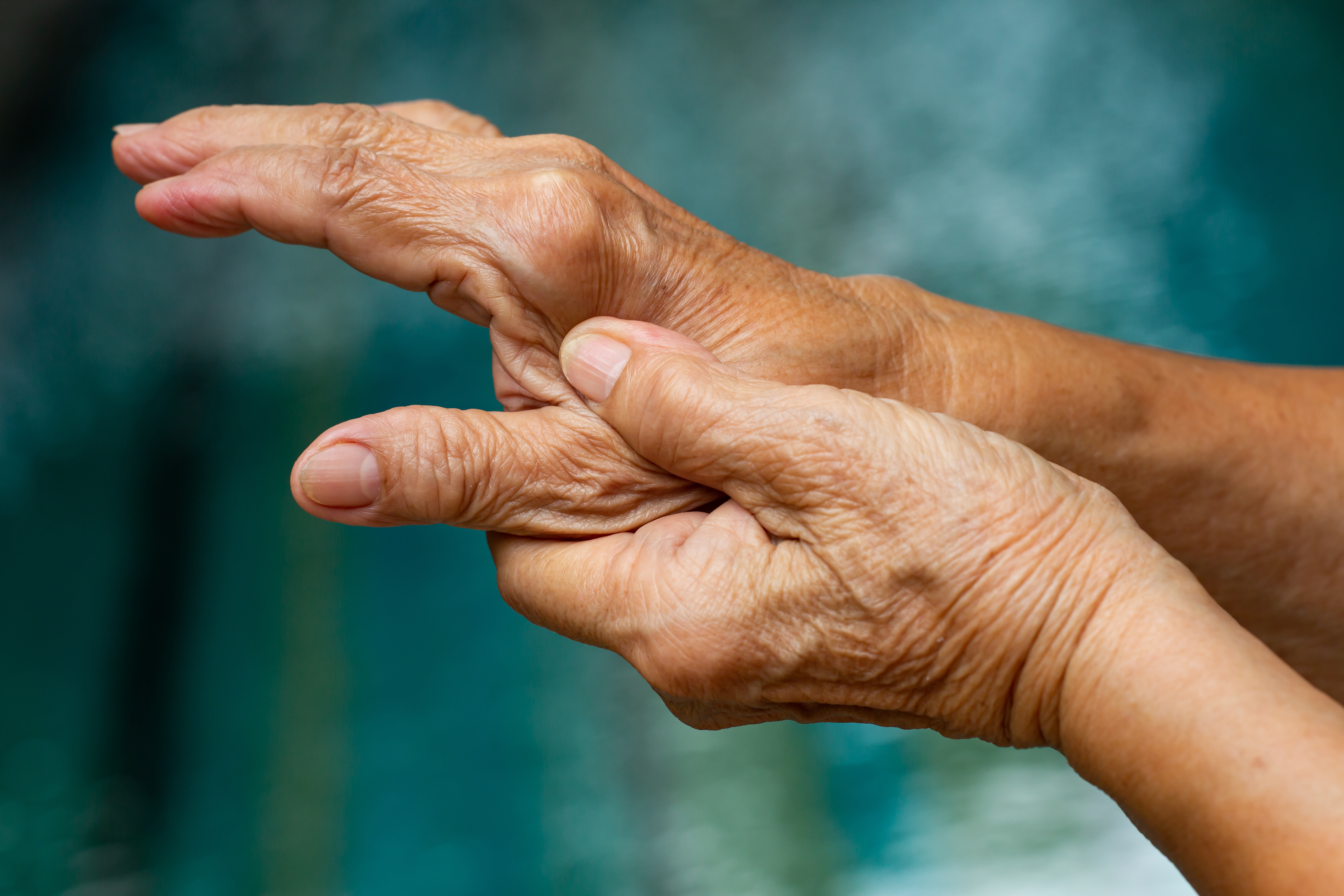
Introduction to Rheumatoid Arthritis and Cartilage Damage
Rheumatoid arthritis (RA) is a complex autoimmune disease that impacts the joints, leading to debilitating pain and reduced quality of life. One of the early indicators of joint damage in RA is the deterioration of articular cartilage, particularly in the metacarpophalangeal (MCP) joints. Recent advancements in imaging technology, specifically ultra-high frequency ultrasound (UHF), have opened potential new avenues for assessing cartilage health.1
The Role of Ultra-high Frequency Ultrasound
UHF ultrasound operates at probe frequencies of 30 MHz or higher, allowing for a spatial resolution of just 30 micrometers. This capability may help clinicians to visualize and measure the intricate structures of cartilage, providing both quantitative and semiquantitative assessments.1 The quantitative approach focuses on measuring cartilage thickness, while the semiquantitative method employs scoring systems to evaluate cartilage damage.
Study Overview and Methodology
In a recent study by Zheng et al published in Quantitative Imaging in Medicine and Surgery1, with 110 patients diagnosed with RA and 110 healthy controls, researchers aimed to identify the vulnerable areas of metacarpal head (MH) cartilage and compare the effectiveness of two different semiquantitative scoring systems. The study utilized both a three-grade and a five-grade scoring system to assess cartilage damage, revealing that the five-grade system detected a higher rate of impairment compared to the three-grade system.1
Findings on Cartilage Damage
The findings indicated that cartilage damage was most prevalent in the ulnar side of the transverse sections and the distal side of the longitudinal sections of the metacarpal head. Specifically, the ulnar side exhibited a damage rate of 7.20%, while the distal side showed a rate of 6.30%.1 These results suggest the importance of targeted assessments in identifying specific areas of vulnerability in RA patients.
Implications for Clinical Practice
The five-grade semiquantitative scoring system for UHF demonstrated reliability for detecting MH cartilage damage in RA. The MCP cartilage of the ulnar and distal sides are particularly susceptible to injury. The implications of these findings are significant for both clinicians and patients. By utilizing UHF ultrasound to better visualize the specific areas of cartilage vulnerability, healthcare providers may be able to better tailor treatment plans to mitigate joint damage.1
Conclusion
The assessment of metacarpal head cartilage via ultrahigh frequency ultrasound represents a promising advancement in the detection and management of rheumatoid arthritis. As research continues to evolve, the integration of advanced imaging techniques like ultra-high frequency ultrasound is likely to enhance our understanding of rheumatoid arthritis and its impact on joint health.
This article provides FUJIFILM Sonosite comments on the results from a single study. Results from case studies are not predictive of results in other cases. Results in other cases may vary. At all times, it is the professional responsibility of the practitioner to exercise independent clinical judgment in each particular situation.
1 - Zheng X, Zhu B, Tang Y, Tang X, Li M, Liu B, Qiu L. The semiquantitative assessment of metacarpal head cartilage damage in rheumatoid arthritis via ultrahigh frequency ultrasound. Quant Imaging Med Surg. 2025 Mar 3;15(3):1927-1937. doi: 10.21037/qims-24-1539. Epub 2025 Feb 26. PMID: 40160650; PMCID: PMC11948411. https://pubmed.ncbi.nlm.nih.gov/40160650/