Skip to main content
Search
Search Toggle Button
Select your language
United States
América Latina
Australia
Brasil
Canada
Canada (Français)
Deutschland
España
France
India
Italia
United Kingdom
Россия
简体中文
日本
Menu
Main menu
Products
Ultrasound systems
Sonosite LX
Unmatched frequency range
Sonosite ST
Procedural Partner
Image
Sonosite MT
New
Built to move
Sonosite PX
Flexible hybrid
Sonosite SII-VA
Intelligence, Inspired by You.
Discover Products
Sonosite LX
Sonosite PX
Sonosite ST
Sonosite SII
Sonosite PIV Assist
New
Ultra-High Frequency
New
Ultra-High Frequency
New
Transducers
Accessories
Sonosite Voice Assist
Synapse Synchronicity
Education
Learn
Clinical Images & Videos
Conferences & Society Meetings
Sonosite Institute
Webinars
Educational Resources
Support
Biomed Service Training
Cleaners and Disinfectants
Connectivity
Contact Support
Dealers & Support Staff
FAQ
Product Retirement
Reimbursement
Security
Symbols Glossary
User Documents
Warranty and Service
About
Why Sonosite
Global Health Program
Leadership Team
Press Releases
What is POCUS?
Careers
Blog
Contact Us
Clinical Images & Videos
Breadcrumb
Home
/
Education
/
Clinical Images & Videos
Search
Clinical Specialties
- Any -
Admin
Anesthesiology
BioMed
Cardiology
Cath Lab
Chiropractic
Clinical Educator
Dermatology
EMED
EMS/Air Med
Endocrinology
FP/GP
Gastroenterology
Global Health
Government
Home Health
ICU/CCU
Informatics
Internal Medicine
Interv Rad
Medical Education
MFM/Perinatology
Nephrology
Neurology
Nursing
OB/GYN
Oncology
Opthamology
OR (Shared System)
Orthopedics
Osteopathic Medicine
Pain Mgmt
Pediatrics
Physical Med & Rehab
Physical Therapy
Podiatry
Pulmonary
Purchasing
Radiology
Respiratory
Rheumatology
SIM Center
Sports Medicine
Sports Team
Surgery
Surgery (Breast)
Urgent Care
Urology
Vascular
Vascular Lab
Vascular Surgery
Vein Clinic
FAST RUQ 3
2044
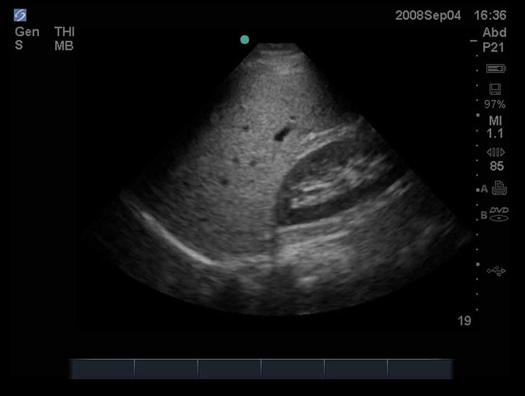
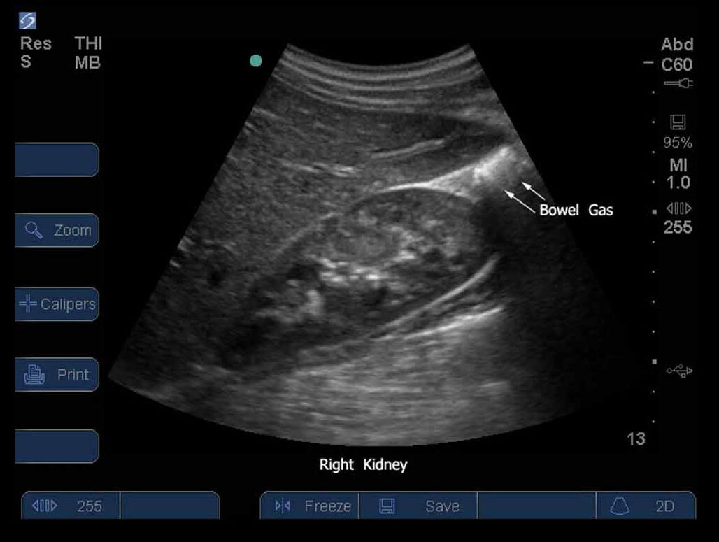
S-Series: Abdomen 02
2116
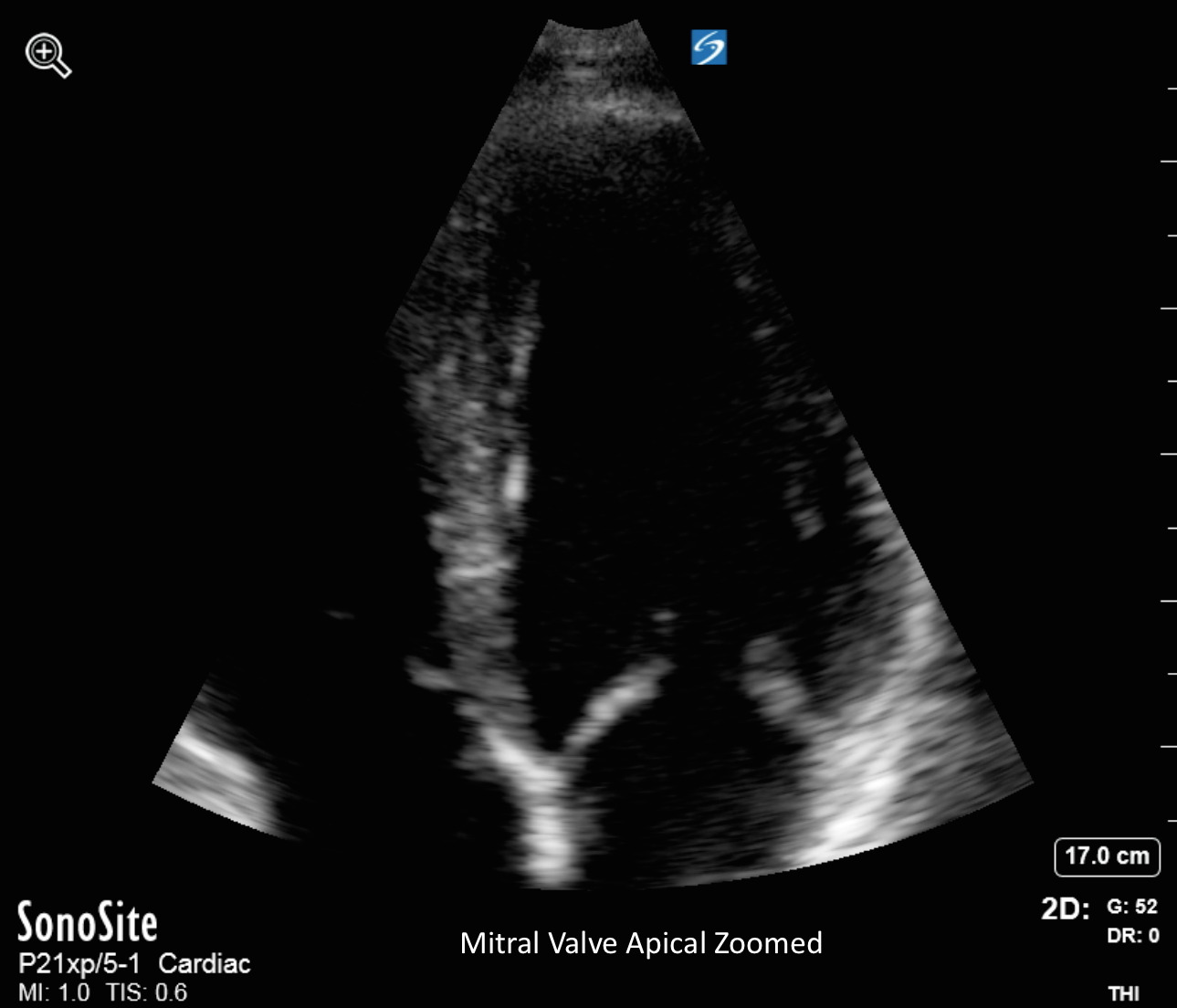
Mitral Valve Apical Zoom
2376
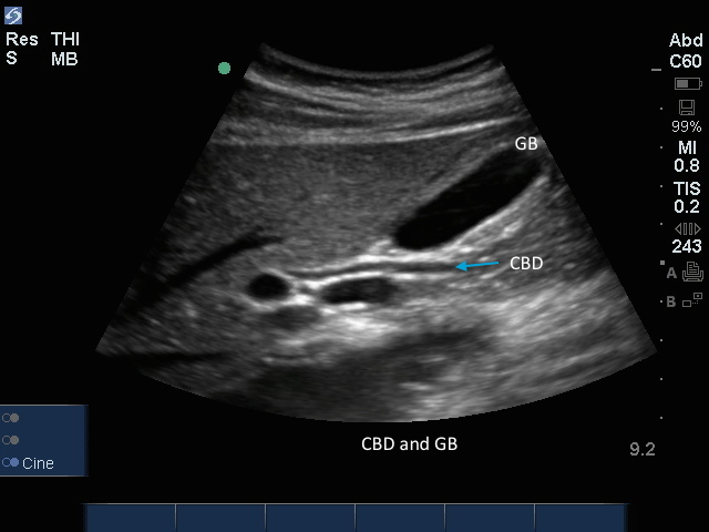
CBD with Gallbladder
2408
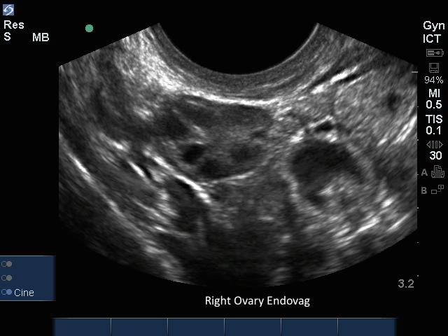
Endovag Rt Ovary
2441
Vascular Access 3
2095
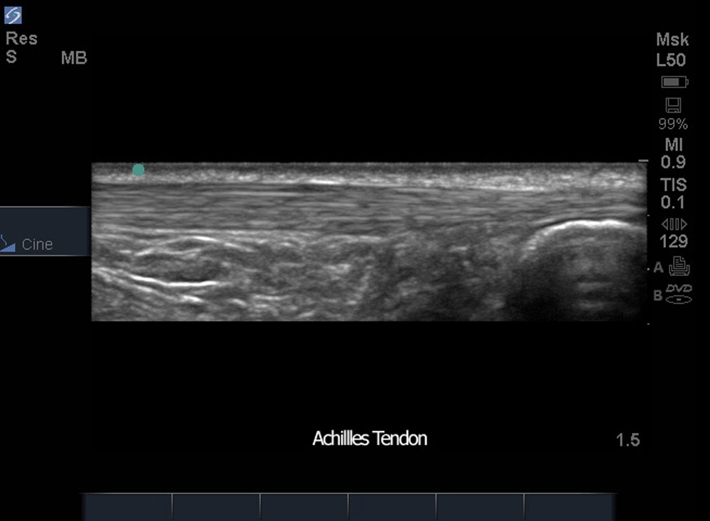
Achilles Tendon
2169
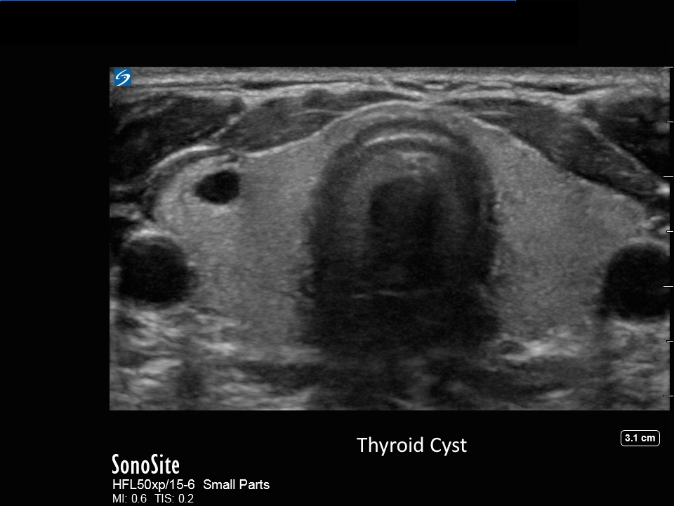
Thyroid Cyst
2387
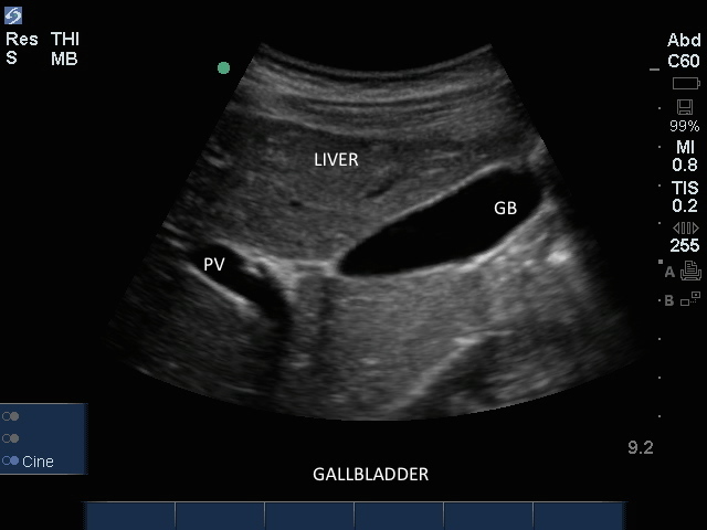
Gallbladder
2420
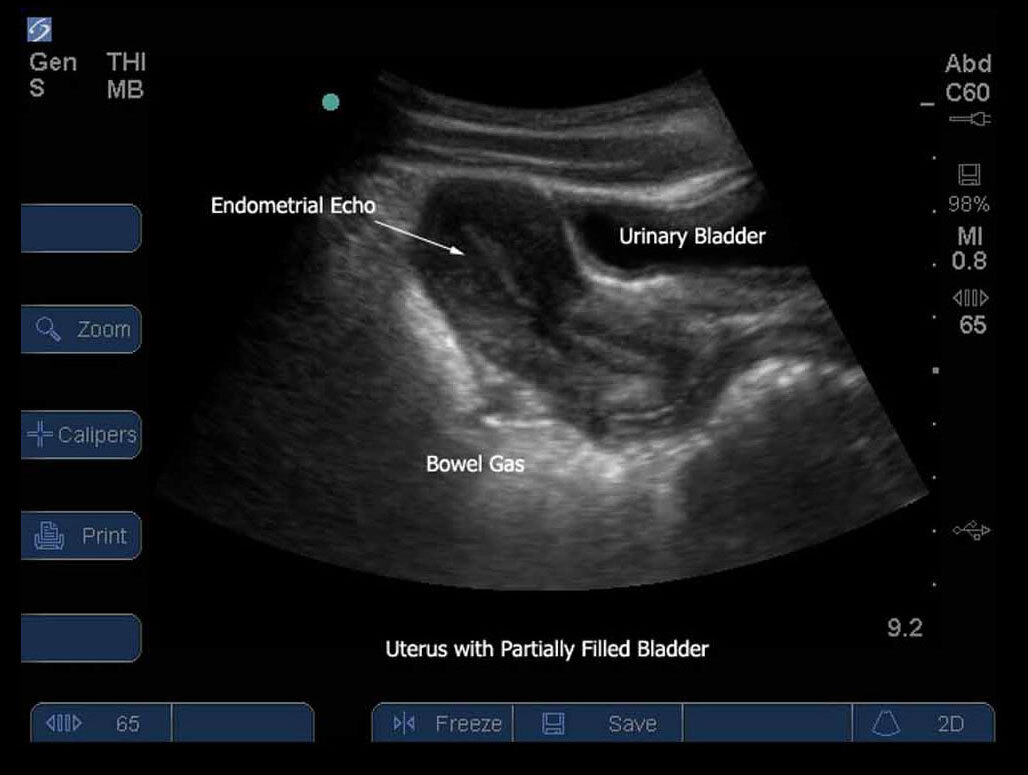
Female Pelvis 11
2106
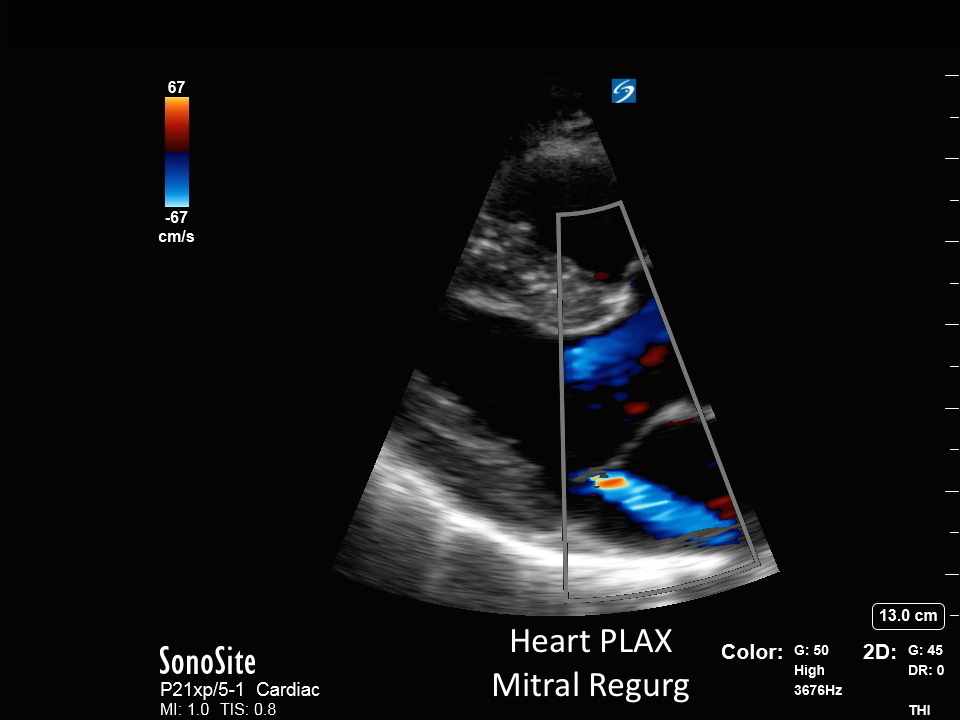
Heart - PLAX Mitral Regurg
2366
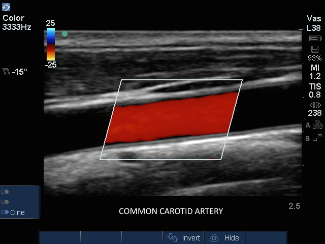
Carotid: CCA Color
2398
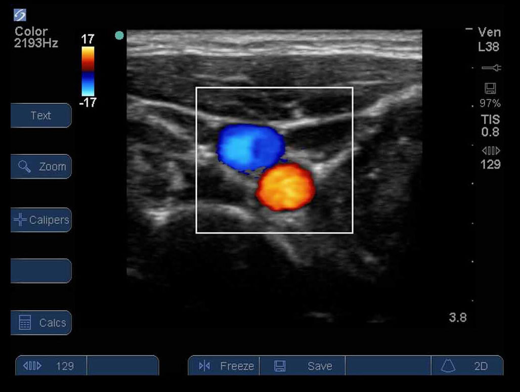
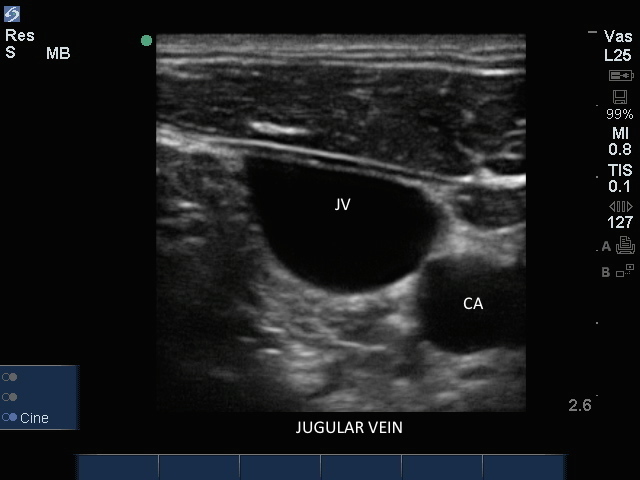
Jugular Vein Color
2431
FAST RUQ 4
2045
S Series: Prox Trv Aorta/Arteries
2117
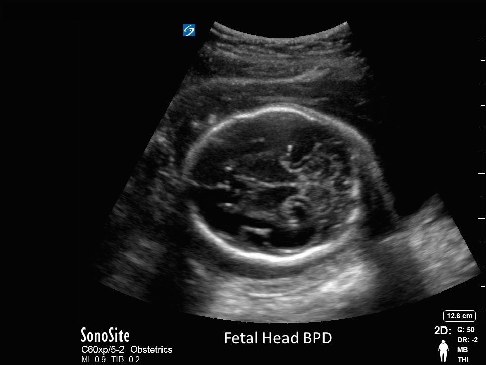
Fetal Head BPD
2377
Pagination
First page
« First
Previous page
‹‹
…
Page
10
Page
11
Page
12
Page
13
Current page
14
Page
15
Page
16
Page
17
Page
18
Next page
››
Last page
Last »
Products
Discover Products
Sonosite MT
Sonosite LX
Sonosite PX
Sonosite ST
Sonosite SII
Sonosite PIV Assist
Ultra-High Frequency
Ultra-High Frequency
Transducers
Accessories
Sonosite Voice Assist
Synapse Synchronicity
Sonosite LX
Education
Learn
Clinical Images & Videos
Conferences & Society Meetings
Sonosite Institute
Webinars
Educational Resources
Support
Biomed Service Training
Cleaners and Disinfectants
Connectivity
Contact Support
Dealers & Support Staff
FAQ
Product Retirement
Reimbursement
Security
Symbols Glossary
User Documents
Warranty and Service
About
Why Sonosite
Global Health Program
Leadership Team
Press Releases
What is POCUS?
Careers
Blog
Contact Us
Select your language
United States
América Latina
Australia
Brasil
Canada
Canada (Français)
Deutschland
España
France
India
Italia
United Kingdom
Россия
简体中文
日本