Primary Care Ultrasound
Primary care physicians are discovering the value of ultrasound visualisation in quickly assessing, diagnosing, and determining treatment options for their patients at the point of care.


Primary care physicians are discovering the value of ultrasound visualisation in quickly assessing, diagnosing, and determining treatment options for their patients at the point of care.
Clinical evidence and research supports using ultrasound as the first diagnostic test for numerous musculoskeletal (MSK) conditions. Diagnostic ultrasound offers a number of important advantages compared to computed tomography (CT) and magnetic resonance imaging (MRI), in terms of safety and effectiveness.
Ultrasound technology provides endocrinologists with tools that can increase diagnostic confidence, improve efficiency, and decrease complications.
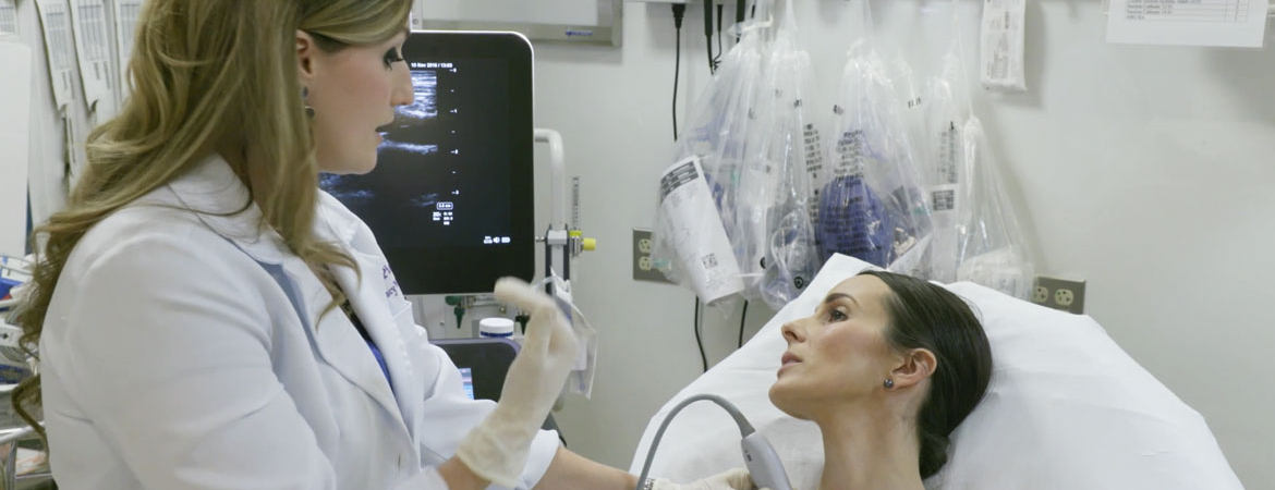
The use of point-of-care ultrasound (POCUS) technology in Women’s Health can increase diagnostic confidence for providers, improve efficiency and decrease complications, therefore reducing costs overall.
You may have noticed that our ultrasound systems include a unique on/off option for needle visualisation. The reason for this relatively insignificant-looking keypad feature leads directly into a very significant Sonosite technology . . . and an explanation for why our needle visualisation software is more advanced than many competitors’.
Let’s start with the necessity for a needle-visualisation on/off switch. The optimal sound waves needed for visualising tissue are different from the optimal sound waves needed for visualising a needle. Consequently—for Sonosite’s advanced needle visualisation—we designed our units to send different pulses for the tissue than for the needle. This patent-pending technology greatly improves needle visualisation compared to targeting the needle with a conventional imaging beam.
When the user doesn’t want to visualise a needle, the optimal thing to do is send only the sound waves intended for the tissue (etc.), not those for the needle. Therefore, we allow the user to turn off that option.
The benefits of Sonosite’s advanced needle visualisation may best be summarised in this concise, albeit rather formal, definition:
Once associated primarily with OB/Gyn, ultrasound continues to grow like a robust, multi-branching tree into evermore areas of medicine. Subscribers to ultrasound news feeds, posts, and journals are regularly presented with new ways of using ultrasound imaging to help improve diagnostic accuracy, boost patient safety, and reduce expenses. Medical schools are starting to add point-of-care ultrasound to their students' must-have list of skills, no matter what their intended specialty
Beyond its use for dynamic scans and on-the-spot assessments, point-of-care ultrasound is being used by physicians in a broad number of fields to help with needle guidance during a variety of procedures. Studies indicate that ultrasound guidance may improve success and decrease complications in central line placements, peripheral vascular access, regional anaesthesia (nerve blocks), lumbar puncture, biopsies, thoracentesis, paracentesis, arthrocentesis, incision and drainage of abscesses, and localization and removal of foreign bodies.
As the list of applications continues to grow, so does the list of medical specialties turning to ultrasound first to help lead the way.
There are places where the price of compromise is too high. Sonosite combines portability and image quality in a unit that is designed to take whatever hospital staff or austere environments dish out.
Orthopaedic surgeons are discovering the value of ultrasound visualisation for assessing injury and function and for improving the accuracy of interventional procedures. Sonosite works closely with orthopaedic surgeons to develop imaging solutions that meet the rapidly expanding clinical requirements of this specialty.