


Exposure to Ionizing Radiation is Growing
每年肾绞痛影响着近120万人口,同时约占住院总量的1%。1 对因肾结石造成肾绞痛患者的诊断往往使用计算机断层扫描(CT)进行,偶尔也会使用静脉泌尿系造影(IVU)。2 虽然CT和IVU检查可以提供准确的诊断信息,精确测量尿酸结石的大小、形状和位置,但他们同样会带来一些负面的效果,3包括将患者暴露在重复电离辐射下。事实上,Joint Comission最近发布了一项关于诊断成像的辐射风险的警告4。报道显示,在过去的20多年时间里,美国人所受电离辐射暴露几乎翻倍。报道指出“… 任何医师可以以任意频率要求患者接受带电离辐射的检查,而往往他们不知道患者此前是否接受过带辐射检查,或此前接受了多少辐射剂量。”
Berrington de Gonzales等综合研究了基于风险模型的2007年美国辐射检查频率,从而估计,由于存在大量的腹部和盆腔检查,在外来约有29,000名癌症病例可能与CT检查有关。5从2006年到2009年的医疗数据显示,CT检查被用于近80%的肾绞痛诊断中。再考虑到肾绞痛经常复发的特点,这进一步加大了患者的辐射相关风险。
Use of Ultrasound to Reduce Risks
与此同时,由于Point-of-Care超声结果可以实时获取,而且毫无辐射。在Joint Commission的警告中,他们推荐减少电离辐射暴露风险的方法之一是使用替代性影像诊断手段,例如超声或者核磁共振,“…如果这一个替代性检查手段可以提供类似水平的诊断信息。”研究发现在绝大部分肾绞痛病例中,超声可以提供可靠的、无创的诊断。6,7
欧洲泌尿学会在其2011年尿石症指南中建议,对于有尿路结石的患者,影像学检查需要在临床检查之后进行。而首选影像学诊断手段是超声:“超声成像应成为主要检查手段。它是一种相对安全(无辐射风险),可重复而且廉价的尿路结石检查方法。”超声检查不仅可以定位在上尿路扩张处的结石,同时也能定位在肾盂、盆腔、肾盂输尿管结合处和膀胱输尿管结合处的结石。8
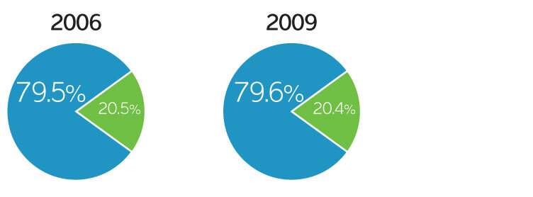
Medicare allowed charges for diagnosing renal colic by type of imaging, 2009 (carrier billed services only)
CT
Ultrasound
Saving money for our nation’s healthcare system
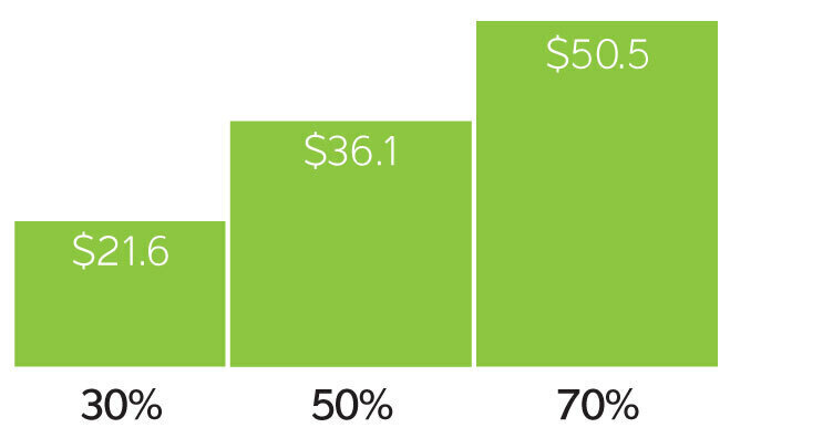
Estimated reductions in 2009 Medicare spending with substitution of diagnostic ultrasound for CT and MRI imaging (in Millions))
除了超声在诊断肾绞痛上的临床效果以外,它相较于CT有显著的成本优势。一份由KNG Health Consulting, LLC9进行的研究显示,如果超声被首先作为诊断工具,将实现全美医疗机构和患者的成本降低。KNG 2009Medicare数据显示如果使用超声替代30%的CT肾绞痛检查,将降低2160万美元成本。如果替代70%,则成本降低将超过5000万美元。总而言之,超声检查不仅是肾绞痛检查安全有效的优先检查手段,同时它还可以让整个医疗系统和患者显著降低医疗成本。
参考资料
1 Wolf JS. Nephrolithiasis: Acute renal colic. Medscape Reference, 2011. Available at: http://emedicine.medscape.com/article/437096-overview
2 Renal colic is a condition that begins as pain in the kidney area or below and radiates through the flank until reaching the bladder. Renal colic pain tends to remain constant and may come in two varieties: dull or acute. Koenig L, Ruiz D, Cornejo A. Potential cost savings from the use of diagnostic ultrasound in the Medicare population. KNG Health Consulting LLC, 2011. Prepared for Sonosite, Inc.
3 Wolf JS. Nephrolithiasis: Acute renal colic. Medscape Reference, 2011. http://emedicine.medscape.com/article/437096-overview
4 The Joint Comission Sentinal Event Alert, issue 47, August 24, 2011. http://www.jointcommission.org/assets/1/18/SEA_471.PDF
5 Berrington de Gonzales A, Mahesh M, Kim K, Bhargavan M, Lewis R, Mettler F, et al. Projected cancers risks from computed Tomographic scans performed in the United States in 2007. Arch Intern Med. 2009;169(22):2071-77.
6 Erwin B, Carroll B, Sommer G. Renal Colic: The role of ultrasound in initial evaluation. Radiology. 1984;(152)147-50.
7 Perven A, Ammar A. Role of ultrasound in evaluation of renal colic and assessment of risk factor for renal calculi. Gomal Journal of Medical Sciences, Jan. - June, 2007;5(1).
8 Turk C, Knoll T, Petrik A, et. al. Guidelines on Urolithiasis, European Association of Urology, 2011.
9 Renal colic is a condition that begins as pain in the kidney area or below and radiates through the flank until reaching the bladder. Renal colic pain tends to remain constant and may come in two varieties: dull or acute. Koenig L, Ruiz D, Cornejo A. Potential cost savings from the use of diagnostic ultrasound in the Medicare population. KNG Health Consulting LLC, 2011. Prepared for Sonosite, Inc.
其他参考资料
Katz SI, Saluja S, Brink JA, et al. Radiation dose associated with unenhanced ct for suspected renal colic: Impact of repetitive studies. Am J Roentgenol. 2006;186(4):1120-4.
Chauhan V, Eskin B, Allegra JR, Cochrane DG. Effect of season, age, and gender on renal colic incidence. Am J Emerg Med. 2004;22(7):560-3
Brenner DJ, Hall EJ. Computed tomography - An increasing source of radiation exposure. New Eng J Med. 2007;357:2277-84.